

138
G. Yang and Y. Huang
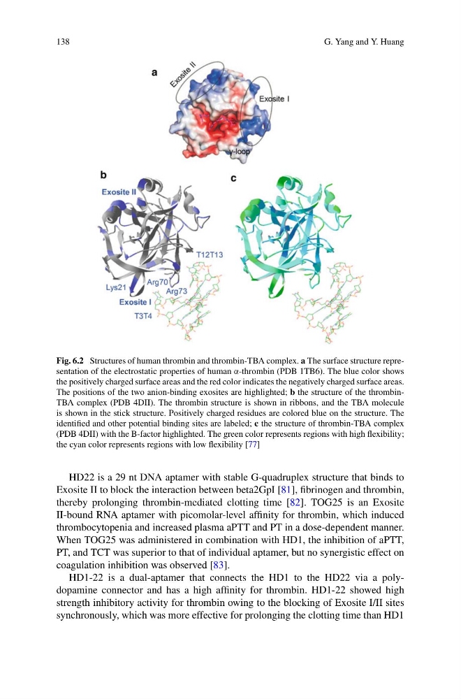
Fig. 6.2 Structures of human thrombin and thrombin-TBA complex. a The surface structure representation of the electrostatic properties of human α-thrombin (PDB 1TB6). The blue color shows
the positively charged surface areas and the red color indicates the negatively charged surface areas.
The positions of the two anion-binding exosites are highlighted; b the structure of the thrombinTBA complex (PDB 4DII). The thrombin structure is shown in ribbons, and the TBA molecule
is shown in the stick structure. Positively charged residues are colored blue on the structure. The
identified and other potential binding sites are labeled; c the structure of thrombin-TBA complex
(PDB 4DII) with the B-factor highlighted. The green color represents regions with high flexibility;
the cyan color represents regions with low flexibility [77]
HD22 is a 29 nt DNA aptamer with stable G-quadruplex structure that binds to
Exosite II to block the interaction between beta2GpI [81], fibrinogen and thrombin,
thereby prolonging thrombin-mediated clotting time [82]. TOG25 is an Exosite
II-bound RNA aptamer with picomolar-level affinity for thrombin, which induced
thrombocytopenia and increased plasma aPTT and PT in a dose-dependent manner.
When TOG25 was administered in combination with HD1, the inhibition of aPTT,
PT, and TCT was superior to that of individual aptamer, but no synergistic effect on
coagulation inhibition was observed [83].
HD1-22 is a dual-aptamer that connects the HD1 to the HD22 via a polydopamine connector and has a high affinity for thrombin. HD1-22 showed high
strength inhibitory activity for thrombin owing to the blocking of Exosite I/II sites
synchronously, which was more effective for prolonging the clotting time than HD1
G. Yang and Y. Huang
Fig. 6.2 Structures of human thrombin and thrombin-TBA complex. a The surface structure representation of the electrostatic properties of human α-thrombin (PDB 1TB6). The blue color shows
the positively charged surface areas and the red color indicates the negatively charged surface areas.
The positions of the two anion-binding exosites are highlighted; b the structure of the thrombinTBA complex (PDB 4DII). The thrombin structure is shown in ribbons, and the TBA molecule
is shown in the stick structure. Positively charged residues are colored blue on the structure. The
identified and other potential binding sites are labeled; c the structure of thrombin-TBA complex
(PDB 4DII) with the B-factor highlighted. The green color represents regions with high flexibility;
the cyan color represents regions with low flexibility [77]
HD22 is a 29 nt DNA aptamer with stable G-quadruplex structure that binds to
Exosite II to block the interaction between beta2GpI [81], fibrinogen and thrombin,
thereby prolonging thrombin-mediated clotting time [82]. TOG25 is an Exosite
II-bound RNA aptamer with picomolar-level affinity for thrombin, which induced
thrombocytopenia and increased plasma aPTT and PT in a dose-dependent manner.
When TOG25 was administered in combination with HD1, the inhibition of aPTT,
PT, and TCT was superior to that of individual aptamer, but no synergistic effect on
coagulation inhibition was observed [83].
HD1-22 is a dual-aptamer that connects the HD1 to the HD22 via a polydopamine connector and has a high affinity for thrombin. HD1-22 showed high
strength inhibitory activity for thrombin owing to the blocking of Exosite I/II sites
synchronously, which was more effective for prolonging the clotting time than HD1