

82
J. Fan et al.
In addition, we improved our sorting algorithm [22] for ordering 2D unarranged points in the skeleton, by introducing a weight-point association to prevent misleading ordering choices in the presence of sharp curvature change.
The results show that the automatic morphology reconstructing method is
reliable and allows to properly describe the morphological evolution of a tendril
after mechanical stimulation.
In the following Sect. 2, we present in details each of the steps involved in the
proposed image-based morphology reconstruction method. Section 3 provides
the analysis and results. And we conclude in Sect. 4 with discussions and future
work.
2 Method
The image-based method we propose for the morphological analysis of tendrils
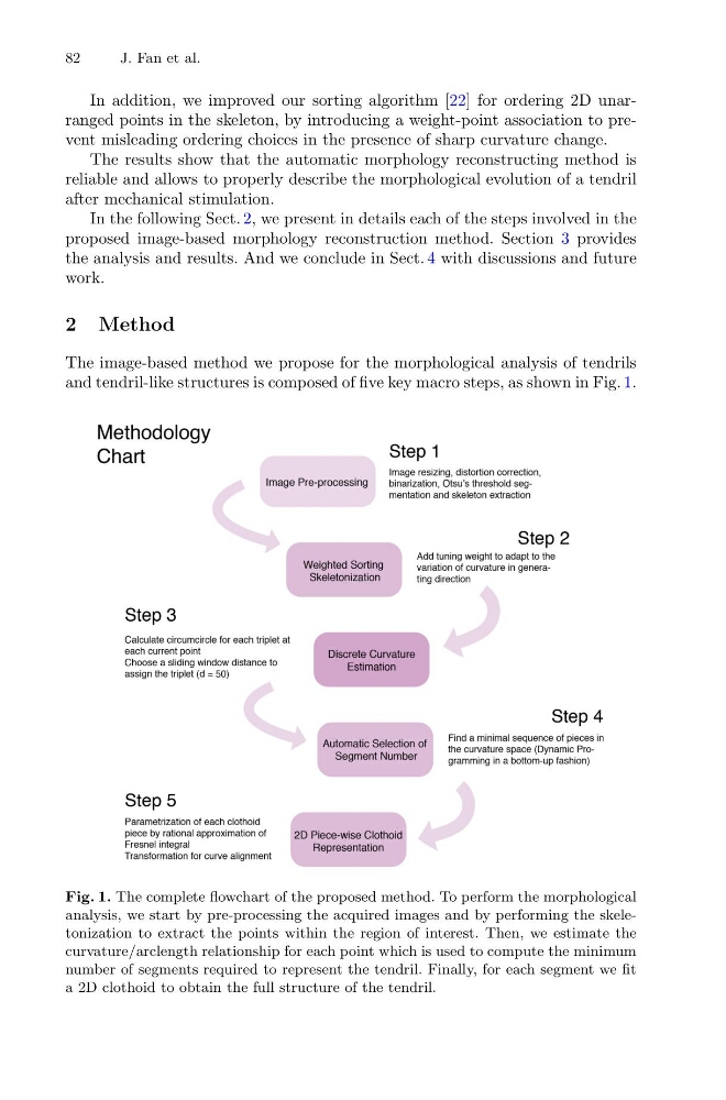
and tendril-like structures is composed of five key macro steps, as shown in Fig. 1.
Fig. 1. The complete flowchart of the proposed method. To perform the morphological
analysis, we start by pre-processing the acquired images and by performing the skeletonization to extract the points within the region of interest. Then, we estimate the
curvature/arclength relationship for each point which is used to compute the minimum
number of segments required to represent the tendril. Finally, for each segment we fit
a 2D clothoid to obtain the full structure of the tendril.
J. Fan et al.
In addition, we improved our sorting algorithm [22] for ordering 2D unarranged points in the skeleton, by introducing a weight-point association to prevent misleading ordering choices in the presence of sharp curvature change.
The results show that the automatic morphology reconstructing method is
reliable and allows to properly describe the morphological evolution of a tendril
after mechanical stimulation.
In the following Sect. 2, we present in details each of the steps involved in the
proposed image-based morphology reconstruction method. Section 3 provides
the analysis and results. And we conclude in Sect. 4 with discussions and future
work.
2 Method
The image-based method we propose for the morphological analysis of tendrils
and tendril-like structures is composed of five key macro steps, as shown in Fig. 1.
Fig. 1. The complete flowchart of the proposed method. To perform the morphological
analysis, we start by pre-processing the acquired images and by performing the skeletonization to extract the points within the region of interest. Then, we estimate the
curvature/arclength relationship for each point which is used to compute the minimum
number of segments required to represent the tendril. Finally, for each segment we fit
a 2D clothoid to obtain the full structure of the tendril.