

Improving Recall in an Associative Neural Network Model of the Hippocampus
3
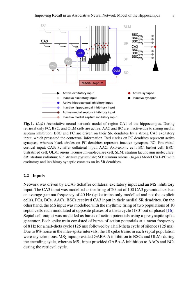
Fig. 1. (Left) Associative neural network model of region CA1 of the hippocampus. During
retrieval only PC, BSC, and OLM cells are active. AAC and BC are inactive due to strong medial
septum inhibition. BSC and PC are driven on their SR dendrites by a strong CA3 excitatory
input, which presented the contextual information. Red circles on PC dendrites represent active
synapses, whereas black circles on PC dendrites represent inactive synapses. EC: Entorhinal
cortical input; CA3: Schaffer collateral input; AAC: Axo-axonic cell; BC: basket cell; BSC:
bistratified cell; OLM: oriens lacunosum-moleculare cell; SLM: stratum lacunosum moleculare;
SR: stratum radiatum; SP: stratum pyramidale; SO: stratum oriens. (Right) Model CA1-PC with
excitatory and inhibitory synaptic contacts on its SR dendrites.
2.2 Inputs
Network was driven by a CA3 Schaffer collateral excitatory input and an MS inhibitory
input. The CA3 input was modelled as the firing of 20 out of 100 CA3 pyramidal cells at
an average gamma frequency of 40 Hz (spike trains only modelled and not the explicit
cells). PCs, BCs, AACs, BSCs received CA3 input in their medial SR dendrites. On the
other hand, the MS input was modelled with the rhythmic firing of two populations of 10
septal cells each modulated at opposite phases of a theta cycle (180° out of phase) [16].
Septal cell output was modelled as bursts of action potentials using a presynaptic spike
generator. Each spike train consisted of bursts of action potentials at a mean frequency
of 8 Hz for a half-theta cycle (125 ms) followed by a half-theta cycle of silence (125 ms).
Due to 8% noise in the inter-spike intervals, the 10 spike trains in each septal population
were asynchronous. MS 2 input provided GABA-A inhibition to BSCs and OLMs during
the encoding cycle, whereas MS 1 input provided GABA-A inhibition to AACs and BCs
during the retrieval cycle.
3
Fig. 1. (Left) Associative neural network model of region CA1 of the hippocampus. During
retrieval only PC, BSC, and OLM cells are active. AAC and BC are inactive due to strong medial
septum inhibition. BSC and PC are driven on their SR dendrites by a strong CA3 excitatory
input, which presented the contextual information. Red circles on PC dendrites represent active
synapses, whereas black circles on PC dendrites represent inactive synapses. EC: Entorhinal
cortical input; CA3: Schaffer collateral input; AAC: Axo-axonic cell; BC: basket cell; BSC:
bistratified cell; OLM: oriens lacunosum-moleculare cell; SLM: stratum lacunosum moleculare;
SR: stratum radiatum; SP: stratum pyramidale; SO: stratum oriens. (Right) Model CA1-PC with
excitatory and inhibitory synaptic contacts on its SR dendrites.
2.2 Inputs
Network was driven by a CA3 Schaffer collateral excitatory input and an MS inhibitory
input. The CA3 input was modelled as the firing of 20 out of 100 CA3 pyramidal cells at
an average gamma frequency of 40 Hz (spike trains only modelled and not the explicit
cells). PCs, BCs, AACs, BSCs received CA3 input in their medial SR dendrites. On the
other hand, the MS input was modelled with the rhythmic firing of two populations of 10
septal cells each modulated at opposite phases of a theta cycle (180° out of phase) [16].
Septal cell output was modelled as bursts of action potentials using a presynaptic spike
generator. Each spike train consisted of bursts of action potentials at a mean frequency
of 8 Hz for a half-theta cycle (125 ms) followed by a half-theta cycle of silence (125 ms).
Due to 8% noise in the inter-spike intervals, the 10 spike trains in each septal population
were asynchronous. MS 2 input provided GABA-A inhibition to BSCs and OLMs during
the encoding cycle, whereas MS 1 input provided GABA-A inhibition to AACs and BCs
during the retrieval cycle.