

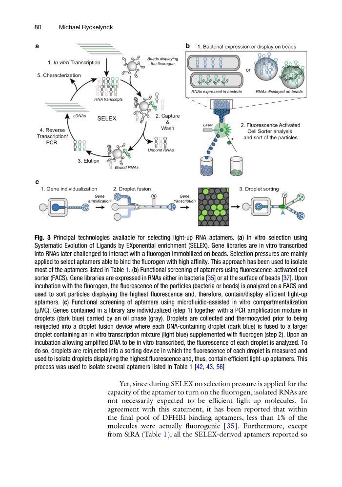
Yet, since during SELEX no selection pressure is applied for the
capacity of the aptamer to turn on the fluorogen, isolated RNAs are
not necessarily expected to be efficient light-up molecules. In
agreement with this statement, it has been reported that within
the final pool of DFHBI-binding aptamers, less than 1% of the
molecules were actually fluorogenic [35]. Furthermore, except
from SiRA (Table 1), all the SELEX-derived aptamers reported so
2. Capture
&
Wash
3. Elution
4. Reverse
Transcription/
PCR
Beads displaying
the fluorogen
1. In vitro Transcription
5. Characterization
cDNAs
RNA transcripts
Unbond RNAs
Bound RNAs
SELEX
a
V
1. Gene individualization
2. Droplet fusion
Gene
amplification
Gene
transcription
c
V
3. Droplet sorting
b
-
+
Laser
2. Fluorescence Activated
Cell Sorter analysis
and sort of the particles
1. Bacterial expression or display on beads
or
RNAs expressed in bacteria
RNAs displayed on beads
Fig. 3 Principal technologies available for selecting light-up RNA aptamers. (a) In vitro selection using
Systematic Evolution of Ligands by EXponential enrichment (SELEX). Gene libraries are in vitro transcribed
into RNAs later challenged to interact with a fluorogen immobilized on beads. Selection pressures are mainly
applied to select aptamers able to bind the fluorogen with high affinity. This approach has been used to isolate
most of the aptamers listed in Table 1. (b) Functional screening of aptamers using fluorescence-activated cell
sorter (FACS). Gene libraries are expressed in RNAs either in bacteria [35] or at the surface of beads [37]. Upon
incubation with the fluorogen, the fluorescence of the particles (bacteria or beads) is analyzed on a FACS and
used to sort particles displaying the highest fluorescence and, therefore, contain/display efficient light-up
aptamers. (c) Functional screening of aptamers using microfluidic-assisted in vitro compartmentalization
(μIVC). Genes contained in a library are individualized (step 1) together with a PCR amplification mixture in
droplets (dark blue) carried by an oil phase (gray). Droplets are collected and thermocycled prior to being
reinjected into a droplet fusion device where each DNA-containing droplet (dark blue) is fused to a larger
droplet containing an in vitro transcription mixture (light blue) supplemented with fluorogen (step 2). Upon an
incubation allowing amplified DNA to be in vitro transcribed, the fluorescence of each droplet is analyzed. To
do so, droplets are reinjected into a sorting device in which the fluorescence of each droplet is measured and
used to isolate droplets displaying the highest fluorescence and, thus, contain efficient light-up aptamers. This
process was used to isolate several aptamers listed in Table 1 [42, 43, 56]
80
Michael Ryckelynck
capacity of the aptamer to turn on the fluorogen, isolated RNAs are
not necessarily expected to be efficient light-up molecules. In
agreement with this statement, it has been reported that within
the final pool of DFHBI-binding aptamers, less than 1% of the
molecules were actually fluorogenic [35]. Furthermore, except
from SiRA (Table 1), all the SELEX-derived aptamers reported so
2. Capture
&
Wash
3. Elution
4. Reverse
Transcription/
PCR
Beads displaying
the fluorogen
1. In vitro Transcription
5. Characterization
cDNAs
RNA transcripts
Unbond RNAs
Bound RNAs
SELEX
a
V
1. Gene individualization
2. Droplet fusion
Gene
amplification
Gene
transcription
c
V
3. Droplet sorting
b
-
+
Laser
2. Fluorescence Activated
Cell Sorter analysis
and sort of the particles
1. Bacterial expression or display on beads
or
RNAs expressed in bacteria
RNAs displayed on beads
Fig. 3 Principal technologies available for selecting light-up RNA aptamers. (a) In vitro selection using
Systematic Evolution of Ligands by EXponential enrichment (SELEX). Gene libraries are in vitro transcribed
into RNAs later challenged to interact with a fluorogen immobilized on beads. Selection pressures are mainly
applied to select aptamers able to bind the fluorogen with high affinity. This approach has been used to isolate
most of the aptamers listed in Table 1. (b) Functional screening of aptamers using fluorescence-activated cell
sorter (FACS). Gene libraries are expressed in RNAs either in bacteria [35] or at the surface of beads [37]. Upon
incubation with the fluorogen, the fluorescence of the particles (bacteria or beads) is analyzed on a FACS and
used to sort particles displaying the highest fluorescence and, therefore, contain/display efficient light-up
aptamers. (c) Functional screening of aptamers using microfluidic-assisted in vitro compartmentalization
(μIVC). Genes contained in a library are individualized (step 1) together with a PCR amplification mixture in
droplets (dark blue) carried by an oil phase (gray). Droplets are collected and thermocycled prior to being
reinjected into a droplet fusion device where each DNA-containing droplet (dark blue) is fused to a larger
droplet containing an in vitro transcription mixture (light blue) supplemented with fluorogen (step 2). Upon an
incubation allowing amplified DNA to be in vitro transcribed, the fluorescence of each droplet is analyzed. To
do so, droplets are reinjected into a sorting device in which the fluorescence of each droplet is measured and
used to isolate droplets displaying the highest fluorescence and, thus, contain efficient light-up aptamers. This
process was used to isolate several aptamers listed in Table 1 [42, 43, 56]
80
Michael Ryckelynck