

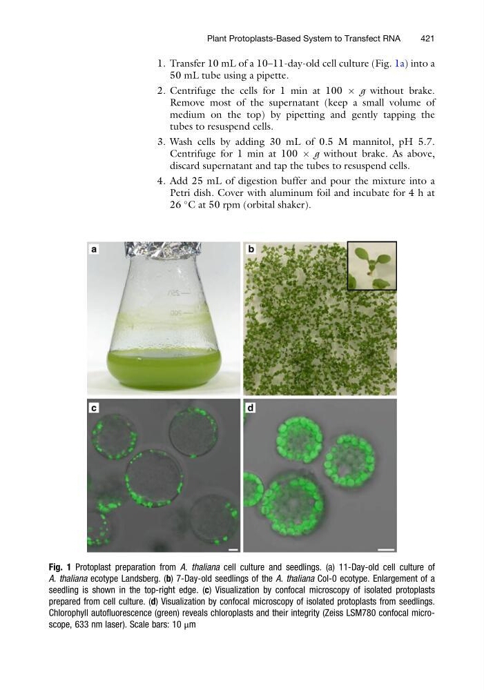
1. Transfer 10 mL of a 10–11-day-old cell culture (Fig. 1a) into a
50 mL tube using a pipette.
2. Centrifuge the cells for 1 min at 100 Â g without brake.
Remove most of the supernatant (keep a small volume of
medium on the top) by pipetting and gently tapping the
tubes to resuspend cells.
3. Wash cells by adding 30 mL of 0.5 M mannitol, pH 5.7.
Centrifuge for 1 min at 100 Â g without brake. As above,
discard supernatant and tap the tubes to resuspend cells.
4. Add 25 mL of digestion buffer and pour the mixture into a
Petri dish. Cover with aluminum foil and incubate for 4 h at
26
C at 50 rpm (orbital shaker).
Fig. 1 Protoplast preparation from A. thaliana cell culture and seedlings. (a) 11-Day-old cell culture of
A. thaliana ecotype Landsberg. (b) 7-Day-old seedlings of the A. thaliana Col-0 ecotype. Enlargement of a
seedling is shown in the top-right edge. (c) Visualization by confocal microscopy of isolated protoplasts
prepared from cell culture. (d) Visualization by confocal microscopy of isolated protoplasts from seedlings.
Chlorophyll autofluorescence (green) reveals chloroplasts and their integrity (Zeiss LSM780 confocal microscope, 633 nm laser). Scale bars: 10 μm
Plant Protoplasts-Based System to Transfect RNA
421
50 mL tube using a pipette.
2. Centrifuge the cells for 1 min at 100 Â g without brake.
Remove most of the supernatant (keep a small volume of
medium on the top) by pipetting and gently tapping the
tubes to resuspend cells.
3. Wash cells by adding 30 mL of 0.5 M mannitol, pH 5.7.
Centrifuge for 1 min at 100 Â g without brake. As above,
discard supernatant and tap the tubes to resuspend cells.
4. Add 25 mL of digestion buffer and pour the mixture into a
Petri dish. Cover with aluminum foil and incubate for 4 h at
26
C at 50 rpm (orbital shaker).
Fig. 1 Protoplast preparation from A. thaliana cell culture and seedlings. (a) 11-Day-old cell culture of
A. thaliana ecotype Landsberg. (b) 7-Day-old seedlings of the A. thaliana Col-0 ecotype. Enlargement of a
seedling is shown in the top-right edge. (c) Visualization by confocal microscopy of isolated protoplasts
prepared from cell culture. (d) Visualization by confocal microscopy of isolated protoplasts from seedlings.
Chlorophyll autofluorescence (green) reveals chloroplasts and their integrity (Zeiss LSM780 confocal microscope, 633 nm laser). Scale bars: 10 μm
Plant Protoplasts-Based System to Transfect RNA
421