

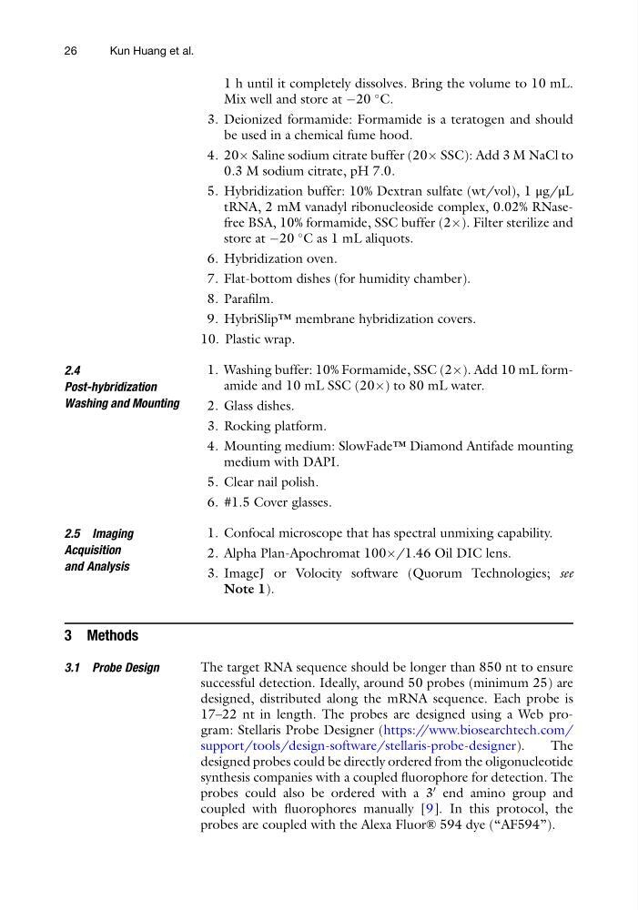
1 h until it completely dissolves. Bring the volume to 10 mL.
Mix well and store at À20
C.
3. Deionized formamide: Formamide is a teratogen and should
be used in a chemical fume hood.
4. 20Â Saline sodium citrate buffer (20Â SSC): Add 3 M NaCl to
0.3 M sodium citrate, pH 7.0.
5. Hybridization buffer: 10% Dextran sulfate (wt/vol), 1 μg/μL
tRNA, 2 mM vanadyl ribonucleoside complex, 0.02% RNasefree BSA, 10% formamide, SSC buffer (2Â). Filter sterilize and
store at À20
C as 1 mL aliquots.
6. Hybridization oven.
7. Flat-bottom dishes (for humidity chamber).
8. Parafilm.
9. HybriSlip™ membrane hybridization covers.
10. Plastic wrap.
2.4
Post-hybridization
Washing and Mounting
1. Washing buffer: 10% Formamide, SSC (2Â). Add 10 mL formamide and 10 mL SSC (20Â) to 80 mL water.
2. Glass dishes.
3. Rocking platform.
4. Mounting medium: SlowFade™ Diamond Antifade mounting
medium with DAPI.
5. Clear nail polish.
6. #1.5 Cover glasses.
2.5 Imaging
Acquisition
and Analysis
1. Confocal microscope that has spectral unmixing capability.
2. Alpha Plan-Apochromat 100Â/1.46 Oil DIC lens.
3. ImageJ or Volocity software (Quorum Technologies; see
Note 1).
3 Methods
3.1 Probe Design
The target RNA sequence should be longer than 850 nt to ensure
successful detection. Ideally, around 50 probes (minimum 25) are
designed, distributed along the mRNA sequence. Each probe is
17–22 nt in length. The probes are designed using a Web program: Stellaris Probe Designer (https://www.biosearchtech.com/
support/tools/design-software/stellaris-probe-designer).
The
designed probes could be directly ordered from the oligonucleotide
synthesis companies with a coupled fluorophore for detection. The
probes could also be ordered with a 3
0 end amino group and
coupled with fluorophores manually [9]. In this protocol, the
probes are coupled with the Alexa Fluor® 594 dye (“AF594”).
26
Kun Huang et al.
Mix well and store at À20
C.
3. Deionized formamide: Formamide is a teratogen and should
be used in a chemical fume hood.
4. 20Â Saline sodium citrate buffer (20Â SSC): Add 3 M NaCl to
0.3 M sodium citrate, pH 7.0.
5. Hybridization buffer: 10% Dextran sulfate (wt/vol), 1 μg/μL
tRNA, 2 mM vanadyl ribonucleoside complex, 0.02% RNasefree BSA, 10% formamide, SSC buffer (2Â). Filter sterilize and
store at À20
C as 1 mL aliquots.
6. Hybridization oven.
7. Flat-bottom dishes (for humidity chamber).
8. Parafilm.
9. HybriSlip™ membrane hybridization covers.
10. Plastic wrap.
2.4
Post-hybridization
Washing and Mounting
1. Washing buffer: 10% Formamide, SSC (2Â). Add 10 mL formamide and 10 mL SSC (20Â) to 80 mL water.
2. Glass dishes.
3. Rocking platform.
4. Mounting medium: SlowFade™ Diamond Antifade mounting
medium with DAPI.
5. Clear nail polish.
6. #1.5 Cover glasses.
2.5 Imaging
Acquisition
and Analysis
1. Confocal microscope that has spectral unmixing capability.
2. Alpha Plan-Apochromat 100Â/1.46 Oil DIC lens.
3. ImageJ or Volocity software (Quorum Technologies; see
Note 1).
3 Methods
3.1 Probe Design
The target RNA sequence should be longer than 850 nt to ensure
successful detection. Ideally, around 50 probes (minimum 25) are
designed, distributed along the mRNA sequence. Each probe is
17–22 nt in length. The probes are designed using a Web program: Stellaris Probe Designer (https://www.biosearchtech.com/
support/tools/design-software/stellaris-probe-designer).
The
designed probes could be directly ordered from the oligonucleotide
synthesis companies with a coupled fluorophore for detection. The
probes could also be ordered with a 3
0 end amino group and
coupled with fluorophores manually [9]. In this protocol, the
probes are coupled with the Alexa Fluor® 594 dye (“AF594”).
26
Kun Huang et al.