

2. Dilute the required amount of the BP stock solution 1:50 in
prewarmed media to generate a 10 mM working stock. Immediately add to cell plates (except to ÀBP controls) 50 μL of the
10 mM BP working stock per 1 mL of media for a final
concentration of 500 μM BP. Gently swirl and return plates
to the incubator for 30 min.
3. During the 30-min BP incubation: Dilute the 16% PFA to 4%
in PBS (use 10Â PBS to account for the 16% PFA volume).
Dilute hydrogen peroxide (most come as 30% wt/wt, which is
~10 M) to 100 mM in PBS. Dissolve sodium ascorbate to 1 M
(in water) and Trolox to 500 mM (in DMSO, sonicate if
necessary). Prepare fresh quencher solution (PBS supplemented with 10 mM sodium ascorbate, 5 mM Trolox, and 10 mM
sodium azide).
4. Bring the plates from the incubator to the bench, add the
100 mM hydrogen peroxide stock into growth medium for a
final concentration of 1 mM (except for the controls without
hydrogen peroxide), swirl, and incubate for 1 min. Proceed
immediately, and shorten rather than extend this incubation
time to minimize the labeling time window.
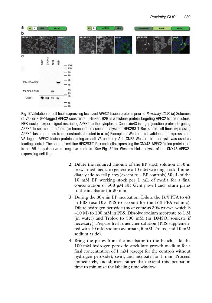
Fig. 2 Validation of cell lines expressing localized APEX2-fusion proteins prior to Proximity-CLIP. (a) Schemes
of V5- or EGFP-tagged APEX2 constructs. L-linker, H2B is a histone protein targeting APEX2 to the nucleus,
NES-nuclear export signal restricting APEX2 to the cytoplasm, Connexin43 is a gap junction protein targeting
APEX2 to cell-cell interface. (b) Immunofluorescence analysis of HEK293 T-Rex stable cell lines expressing
APEX2-fusion proteins from constructs depicted in a. (c) Example of Western blot validation of expression of
V5-tagged APEX2-fusion proteins, using an anti-V5 antibody. Anti-CNBP Western blot analysis was used as
loading control. The parental cell line HEK293 T-Rex and cells expressing the CNX43-APEX2 fusion protein that
is not V5-tagged serve as negative controls. See Fig. 3f for Western blot analysis of the CNX43-APEX2expressing cell line
Proximity-CLIP
289
prewarmed media to generate a 10 mM working stock. Immediately add to cell plates (except to ÀBP controls) 50 μL of the
10 mM BP working stock per 1 mL of media for a final
concentration of 500 μM BP. Gently swirl and return plates
to the incubator for 30 min.
3. During the 30-min BP incubation: Dilute the 16% PFA to 4%
in PBS (use 10Â PBS to account for the 16% PFA volume).
Dilute hydrogen peroxide (most come as 30% wt/wt, which is
~10 M) to 100 mM in PBS. Dissolve sodium ascorbate to 1 M
(in water) and Trolox to 500 mM (in DMSO, sonicate if
necessary). Prepare fresh quencher solution (PBS supplemented with 10 mM sodium ascorbate, 5 mM Trolox, and 10 mM
sodium azide).
4. Bring the plates from the incubator to the bench, add the
100 mM hydrogen peroxide stock into growth medium for a
final concentration of 1 mM (except for the controls without
hydrogen peroxide), swirl, and incubate for 1 min. Proceed
immediately, and shorten rather than extend this incubation
time to minimize the labeling time window.
Fig. 2 Validation of cell lines expressing localized APEX2-fusion proteins prior to Proximity-CLIP. (a) Schemes
of V5- or EGFP-tagged APEX2 constructs. L-linker, H2B is a histone protein targeting APEX2 to the nucleus,
NES-nuclear export signal restricting APEX2 to the cytoplasm, Connexin43 is a gap junction protein targeting
APEX2 to cell-cell interface. (b) Immunofluorescence analysis of HEK293 T-Rex stable cell lines expressing
APEX2-fusion proteins from constructs depicted in a. (c) Example of Western blot validation of expression of
V5-tagged APEX2-fusion proteins, using an anti-V5 antibody. Anti-CNBP Western blot analysis was used as
loading control. The parental cell line HEK293 T-Rex and cells expressing the CNX43-APEX2 fusion protein that
is not V5-tagged serve as negative controls. See Fig. 3f for Western blot analysis of the CNX43-APEX2expressing cell line
Proximity-CLIP
289