

3. Equilibrate Whatman paper and nitrocellulose membrane
(8.0 Â 6.5 cm each) in transfer buffer (do not soak the gel in
transfer buffer).
4. Remove the wells of the gel using a razor blade.
5. Transfer the gel onto the nitrocellulose membrane. Therefore,
place a pre-soaked Whatman paper on top of the gel. Remove
the air bubbles and turn the plate over. Place the pre-soaked
membrane on the gel. Again, remove the air bubbles and add a
pre-soaked Whatman paper on top.
6. Fill the inner chamber with transfer buffer containing 10%
methanol and the outer chamber with cold ddH 2 O.
7. Transfer for 1 h at 30 V.
8. After blotting, disassemble the XCell II Blot Module and measure radioactivity in the gel and the membrane to verify efficient
transfer.
9. Rinse the membrane in 1Â PBS buffer.
10. Wrap membrane in saran wrap and place it into an X-ray
cassette along with a fluorescent ruler.
11. Expose the membrane to an X-ray film at À80
C for 3 h or
overnight and develop the film in the darkroom using standard
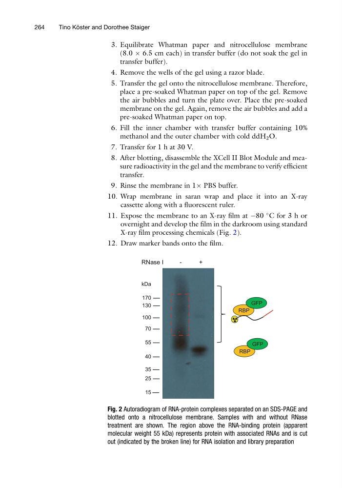
X-ray film processing chemicals (Fig. 2).
12. Draw marker bands onto the film.
170
130
100
70
55
40
35
25
15
RBP
GFP
RBP
GFP
RNase I
-
+
kDa
Fig. 2 Autoradiogram of RNA-protein complexes separated on an SDS-PAGE and
blotted onto a nitrocellulose membrane. Samples with and without RNase
treatment are shown. The region above the RNA-binding protein (apparent
molecular weight 55 kDa) represents protein with associated RNAs and is cut
out (indicated by the broken line) for RNA isolation and library preparation
264
Tino Ko ¨ ster and Dorothee Staiger
(8.0 Â 6.5 cm each) in transfer buffer (do not soak the gel in
transfer buffer).
4. Remove the wells of the gel using a razor blade.
5. Transfer the gel onto the nitrocellulose membrane. Therefore,
place a pre-soaked Whatman paper on top of the gel. Remove
the air bubbles and turn the plate over. Place the pre-soaked
membrane on the gel. Again, remove the air bubbles and add a
pre-soaked Whatman paper on top.
6. Fill the inner chamber with transfer buffer containing 10%
methanol and the outer chamber with cold ddH 2 O.
7. Transfer for 1 h at 30 V.
8. After blotting, disassemble the XCell II Blot Module and measure radioactivity in the gel and the membrane to verify efficient
transfer.
9. Rinse the membrane in 1Â PBS buffer.
10. Wrap membrane in saran wrap and place it into an X-ray
cassette along with a fluorescent ruler.
11. Expose the membrane to an X-ray film at À80
C for 3 h or
overnight and develop the film in the darkroom using standard
X-ray film processing chemicals (Fig. 2).
12. Draw marker bands onto the film.
170
130
100
70
55
40
35
25
15
RBP
GFP
RBP
GFP
RNase I
-
+
kDa
Fig. 2 Autoradiogram of RNA-protein complexes separated on an SDS-PAGE and
blotted onto a nitrocellulose membrane. Samples with and without RNase
treatment are shown. The region above the RNA-binding protein (apparent
molecular weight 55 kDa) represents protein with associated RNAs and is cut
out (indicated by the broken line) for RNA isolation and library preparation
264
Tino Ko ¨ ster and Dorothee Staiger