

the last Csy4∗ codon, a glycine codon is inserted and then a
SV40 nuclear localization signal flanked by two in-frame KpnI
sites: 5
0 -GGA-GGTACC-CCTAAGAAAAAGCGTAAGGTTGGTACC-3
0 . The entire cassette is flanked by Gateway attB1
and attB2 sites at the 5
0 and 3
0 ends, respectively (attB1Csy4∗-KpnI-NLS-KpnI-attB2).
2. Gateway BP Clonase II (Invitrogen).
3. Gateway LR Clonase II (Invitrogen).
4. pDONR221 Gateway donor vector (Invitrogen).
5. pGWB405 destination vector (Addgene #74799, [17]).
6. KpnI restriction enzyme and 10Â buffer.
7. T4 DNA ligase.
8. Electroporator and electroporation cuvettes with 0.2 cm electrode gap width.
9. Escherichia coli XL1 Blue MRF’ electrocompetent cells.
10. Agrobacterium tumefaciens AGL1 electrocompetent cells.
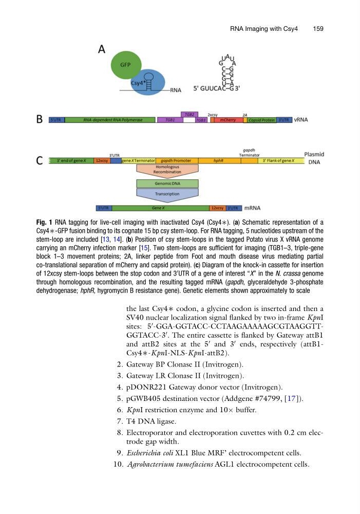
Fig. 1 RNA tagging for live-cell imaging with inactivated Csy4 (Csy4∗). (a) Schematic representation of a
Csy4∗-GFP fusion binding to its cognate 15 bp csy stem-loop. For RNA tagging, 5 nucleotides upstream of the
stem-loop are included [13, 14]. (b) Position of csy stem-loops in the tagged Potato virus X vRNA genome
carrying an mCherry infection marker [15]. Two stem-loops are sufficient for imaging (TGB1–3, triple-gene
block 1–3 movement proteins; 2A, linker peptide from Foot and mouth disease virus mediating partial
co-translational separation of mCherry and capsid protein). (c) Diagram of the knock-in cassette for insertion
of 12xcsy stem-loops between the stop codon and 3
0 UTR of a gene of interest “X” in the N. crassa genome
through homologous recombination, and the resulting tagged mRNA (gapdh, glyceraldehyde 3-phosphate
dehydrogenase; hphR, hygromycin B resistance gene). Genetic elements shown approximately to scale
RNA Imaging with Csy4
159
SV40 nuclear localization signal flanked by two in-frame KpnI
sites: 5
0 -GGA-GGTACC-CCTAAGAAAAAGCGTAAGGTTGGTACC-3
0 . The entire cassette is flanked by Gateway attB1
and attB2 sites at the 5
0 and 3
0 ends, respectively (attB1Csy4∗-KpnI-NLS-KpnI-attB2).
2. Gateway BP Clonase II (Invitrogen).
3. Gateway LR Clonase II (Invitrogen).
4. pDONR221 Gateway donor vector (Invitrogen).
5. pGWB405 destination vector (Addgene #74799, [17]).
6. KpnI restriction enzyme and 10Â buffer.
7. T4 DNA ligase.
8. Electroporator and electroporation cuvettes with 0.2 cm electrode gap width.
9. Escherichia coli XL1 Blue MRF’ electrocompetent cells.
10. Agrobacterium tumefaciens AGL1 electrocompetent cells.
Fig. 1 RNA tagging for live-cell imaging with inactivated Csy4 (Csy4∗). (a) Schematic representation of a
Csy4∗-GFP fusion binding to its cognate 15 bp csy stem-loop. For RNA tagging, 5 nucleotides upstream of the
stem-loop are included [13, 14]. (b) Position of csy stem-loops in the tagged Potato virus X vRNA genome
carrying an mCherry infection marker [15]. Two stem-loops are sufficient for imaging (TGB1–3, triple-gene
block 1–3 movement proteins; 2A, linker peptide from Foot and mouth disease virus mediating partial
co-translational separation of mCherry and capsid protein). (c) Diagram of the knock-in cassette for insertion
of 12xcsy stem-loops between the stop codon and 3
0 UTR of a gene of interest “X” in the N. crassa genome
through homologous recombination, and the resulting tagged mRNA (gapdh, glyceraldehyde 3-phosphate
dehydrogenase; hphR, hygromycin B resistance gene). Genetic elements shown approximately to scale
RNA Imaging with Csy4
159