

mRNA is marked by the hybridization of a labeled antisense RNA
probe, which is used for detection. The labeled antisense probes can
be easily generated by transcription from a PCR product of a gene
of interest in the presence of a small fraction of labeled uracil.
Commonly used labels are digoxigenin, biotin, and fluorescein,
which do not interfere with the RNA polymerase during RNA
synthesis and which do not hinder nucleic acid hybridization.
Afterwards, the bound probe can be visualized by enzyme-coupled
antibodies in the investigated tissues and cells, respectively. The
alkaline phosphatase (AP)-coupled antibody detecting the labeled
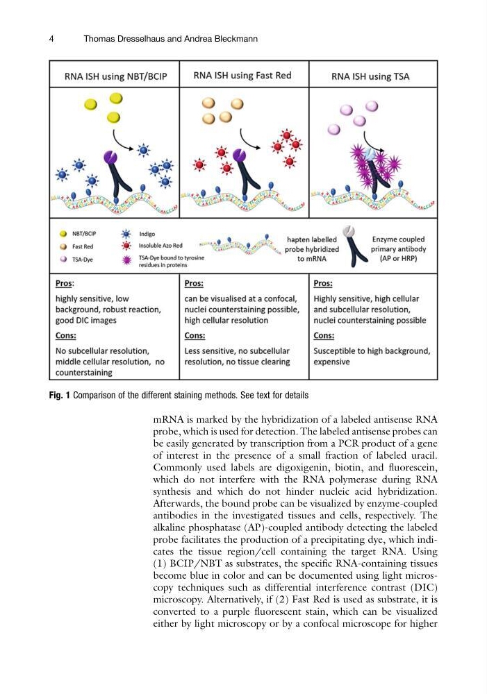
probe facilitates the production of a precipitating dye, which indicates the tissue region/cell containing the target RNA. Using
(1) BCIP/NBT as substrates, the specific RNA-containing tissues
become blue in color and can be documented using light microscopy techniques such as differential interference contrast (DIC)
microscopy. Alternatively, if (2) Fast Red is used as substrate, it is
converted to a purple fluorescent stain, which can be visualized
either by light microscopy or by a confocal microscope for higher
Fig. 1 Comparison of the different staining methods. See text for details
4
Thomas Dresselhaus and Andrea Bleckmann
probe, which is used for detection. The labeled antisense probes can
be easily generated by transcription from a PCR product of a gene
of interest in the presence of a small fraction of labeled uracil.
Commonly used labels are digoxigenin, biotin, and fluorescein,
which do not interfere with the RNA polymerase during RNA
synthesis and which do not hinder nucleic acid hybridization.
Afterwards, the bound probe can be visualized by enzyme-coupled
antibodies in the investigated tissues and cells, respectively. The
alkaline phosphatase (AP)-coupled antibody detecting the labeled
probe facilitates the production of a precipitating dye, which indicates the tissue region/cell containing the target RNA. Using
(1) BCIP/NBT as substrates, the specific RNA-containing tissues
become blue in color and can be documented using light microscopy techniques such as differential interference contrast (DIC)
microscopy. Alternatively, if (2) Fast Red is used as substrate, it is
converted to a purple fluorescent stain, which can be visualized
either by light microscopy or by a confocal microscope for higher
Fig. 1 Comparison of the different staining methods. See text for details
4
Thomas Dresselhaus and Andrea Bleckmann