

3. Observe samples by confocal microscopy. Use a 20Â objective
to locate the area of interest (i.e., the boundary area of the
agroinfiltrated patch) (see Note 9).
4. Use 40Â or 63Â objectives to optimize microscopy settings to
differentiate the following four sample types:
(a) Set I: samples expressing pCAMBIA1390-35S-MS2 FD -
GFP only.
(b) Set II: samples expressing pCAMBIA1390-35S-MS2 FD -
GFP and pCAMBIA1390-35S-FT SL24 .
(c) Set III: samples expressing pCAMBIA1390-35S-MS2 FD -
GFP and pCAMBIA1390-35S-FT (non-tagged mRNA
control).
(d) Set IV: samples expressing pCAMBIA1390-35S-MS2 FD -
GFP and pCAMBIA1390-35S-RFP SL24 (nonmobile
mRNA control).
5. Use 40Â or 63Â objectives to record Z-stack or time-series
images (see Note 10) with support by the microscope software
(e.g., ZEN by Carl Zeiss, Oberkochen, Germany).
6. Process and analyze the acquired images with image processing
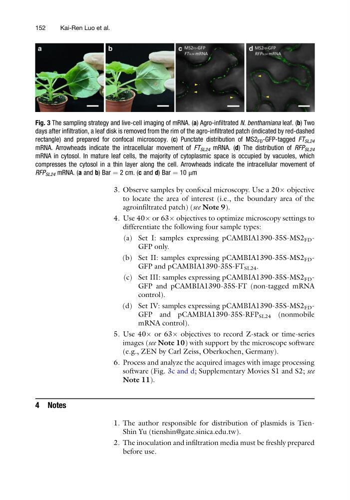
software (Fig. 3c and d; Supplementary Movies S1 and S2; see
Note 11).
4 Notes
1. The author responsible for distribution of plasmids is TienShin Yu (tienshin@gate.sinica.edu.tw).
2. The inoculation and infiltration media must be freshly prepared
before use.
Fig. 3 The sampling strategy and live-cell imaging of mRNA. (a) Agro-infiltrated N. benthamiana leaf. (b) Two
days after infiltration, a leaf disk is removed from the rim of the agro-infiltrated patch (indicated by red-dashed
rectangle) and prepared for confocal microscopy. (c) Punctate distribution of MS2 FD -GFP-tagged FT SL24
mRNA. Arrowheads indicate the intracellular movement of FT SL24 mRNA. (d) The distribution of RFP SL24
mRNA in cytosol. In mature leaf cells, the majority of cytoplasmic space is occupied by vacuoles, which
compresses the cytosol in a thin layer along the cell. Arrowheads indicate the intracellular movement of
RFP SL24 mRNA. (a and b) Bar ¼ 2 cm. (c and d) Bar ¼ 10 μm
152
Kai-Ren Luo et al.
to locate the area of interest (i.e., the boundary area of the
agroinfiltrated patch) (see Note 9).
4. Use 40Â or 63Â objectives to optimize microscopy settings to
differentiate the following four sample types:
(a) Set I: samples expressing pCAMBIA1390-35S-MS2 FD -
GFP only.
(b) Set II: samples expressing pCAMBIA1390-35S-MS2 FD -
GFP and pCAMBIA1390-35S-FT SL24 .
(c) Set III: samples expressing pCAMBIA1390-35S-MS2 FD -
GFP and pCAMBIA1390-35S-FT (non-tagged mRNA
control).
(d) Set IV: samples expressing pCAMBIA1390-35S-MS2 FD -
GFP and pCAMBIA1390-35S-RFP SL24 (nonmobile
mRNA control).
5. Use 40Â or 63Â objectives to record Z-stack or time-series
images (see Note 10) with support by the microscope software
(e.g., ZEN by Carl Zeiss, Oberkochen, Germany).
6. Process and analyze the acquired images with image processing
software (Fig. 3c and d; Supplementary Movies S1 and S2; see
Note 11).
4 Notes
1. The author responsible for distribution of plasmids is TienShin Yu (tienshin@gate.sinica.edu.tw).
2. The inoculation and infiltration media must be freshly prepared
before use.
Fig. 3 The sampling strategy and live-cell imaging of mRNA. (a) Agro-infiltrated N. benthamiana leaf. (b) Two
days after infiltration, a leaf disk is removed from the rim of the agro-infiltrated patch (indicated by red-dashed
rectangle) and prepared for confocal microscopy. (c) Punctate distribution of MS2 FD -GFP-tagged FT SL24
mRNA. Arrowheads indicate the intracellular movement of FT SL24 mRNA. (d) The distribution of RFP SL24
mRNA in cytosol. In mature leaf cells, the majority of cytoplasmic space is occupied by vacuoles, which
compresses the cytosol in a thin layer along the cell. Arrowheads indicate the intracellular movement of
RFP SL24 mRNA. (a and b) Bar ¼ 2 cm. (c and d) Bar ¼ 10 μm
152
Kai-Ren Luo et al.