

system is a reliable strategy to dissect spatial and temporal regulation of mobile mRNAs in real time and under subcellular
resolution.
2 Materials
2.1 Molecular
Cloning
1. The following plasmids are available upon request (see Note 1):
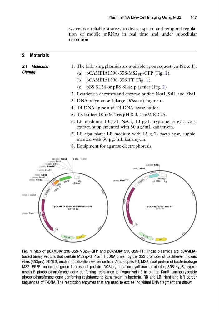
(a) pCAMBIA1390-35S-MS2 FD -GFP (Fig. 1).
(b) pCAMBIA1390-35S-FT (Fig. 1).
(c) pBS-SL24 or pBS-SL48 plasmids (Fig. 2).
2. Restriction enzymes and enzyme buffer: NotI, SalI, and XbaI.
3. DNA polymerase I, large (Klenow) fragment.
4. T4 DNA ligase and T4 DNA ligase buffer.
5. TE buffer: 10 mM Tris pH 8.0, 1 mM EDTA.
6. LB medium: 10 g/L NaCl, 10 g/L tryptone, 5 g/L yeast
extract, supplemented with 50 μg/mL kanamycin.
7. LB agar plate: LB medium with 15 g/L bacto-agar, supplemented with 50 μg/mL kanamycin.
8. Equipment for agarose electrophoresis.
Fig. 1 Map of pCAMBIA1390-35S-MS2 FD -GFP and pCAMBIA1390-35S-FT. These plasmids are pCAMBIAbased binary vectors that contain MS2 FD -GFP or FT cDNA driven by the 35S promoter of cauliflower mosaic
virus (35Spro). FDNLS, nuclear localization sequence from Arabidopsis FD; MS2, coat protein of bacteriophage
MS2; EGFP: enhanced green fluorescent protein; NOSter, nopaline synthase terminator; 35S-HygR, hygromycin B phosphotransferase gene conferring resistance to hygromycin B in plants; KanR, aminoglycoside
phosphotransferase gene conferring resistance to kanamycin in bacteria. RB and LB, right and left border
sequences of T-DNA. The restriction enzymes that are used to excise individual DNA fragment are shown
Plant mRNA Live-Cell Imaging Using MS2
147
resolution.
2 Materials
2.1 Molecular
Cloning
1. The following plasmids are available upon request (see Note 1):
(a) pCAMBIA1390-35S-MS2 FD -GFP (Fig. 1).
(b) pCAMBIA1390-35S-FT (Fig. 1).
(c) pBS-SL24 or pBS-SL48 plasmids (Fig. 2).
2. Restriction enzymes and enzyme buffer: NotI, SalI, and XbaI.
3. DNA polymerase I, large (Klenow) fragment.
4. T4 DNA ligase and T4 DNA ligase buffer.
5. TE buffer: 10 mM Tris pH 8.0, 1 mM EDTA.
6. LB medium: 10 g/L NaCl, 10 g/L tryptone, 5 g/L yeast
extract, supplemented with 50 μg/mL kanamycin.
7. LB agar plate: LB medium with 15 g/L bacto-agar, supplemented with 50 μg/mL kanamycin.
8. Equipment for agarose electrophoresis.
Fig. 1 Map of pCAMBIA1390-35S-MS2 FD -GFP and pCAMBIA1390-35S-FT. These plasmids are pCAMBIAbased binary vectors that contain MS2 FD -GFP or FT cDNA driven by the 35S promoter of cauliflower mosaic
virus (35Spro). FDNLS, nuclear localization sequence from Arabidopsis FD; MS2, coat protein of bacteriophage
MS2; EGFP: enhanced green fluorescent protein; NOSter, nopaline synthase terminator; 35S-HygR, hygromycin B phosphotransferase gene conferring resistance to hygromycin B in plants; KanR, aminoglycoside
phosphotransferase gene conferring resistance to kanamycin in bacteria. RB and LB, right and left border
sequences of T-DNA. The restriction enzymes that are used to excise individual DNA fragment are shown
Plant mRNA Live-Cell Imaging Using MS2
147