

48
R. W. Taylor and V. Sandoghdar
(a)
(ii)
(iii)
(b)
Empty
Full
(i)
(i)
(ii)
(iii)
(iv)
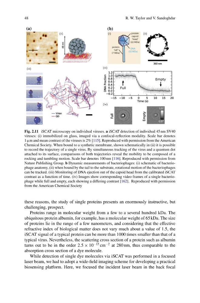
Fig. 2.11 iSCAT microscopy on individual viruses. a iSCAT detection of individual 45 nm SV40
viruses: (i) immobilized on glass, imaged via a confocal-reflection modality. Scale bar denotes
1 µm and mean contrast of the viruses is 2% [115]. Reproduced with permission from the American
Chemical Society. When bound to a synthetic membrane, shown schematically in (ii) it is possible
to record the trajectory of a single virus. By simultaneous tracking of the virus and a quantum dot
attached to its surface, comparisons of both trajectories reveal the mobility to be composed of a
rocking and tumbling motion. Scale bar denotes 100 nm [116]. Reproduced with permission from
Nature Publishing Group. b Dynamic measurements of bacteriophages: (i) schematic of bacteriophage anatomy, (ii) when bound by the tail to the substrate, rotational motion of the bacteriophages
can be tracked. (iii) Monitoring of DNA ejection out of the capsid head from the calibrated iSCAT
contrast as a function of time. (iv) Images show corresponding video frames of a single bacteriophage while full and empty, each showing a differing contrast [162]. Reproduced with permission
from the American Chemical Society
these reasons, the study of single proteins presents an enormously instructive, but
challenging, prospect.
Proteins range in molecular weight from a few to a several hundred kDa. The
ubiquitous protein albumin, for example, has a molecular weight of 65 kDa. The size
of proteins lie in the range of a few nanometers, and considering that the effective
refractive index of biological matter does not vary much about a value of 1.5, the
iSCAT signal of a typical protein can be more than 1000 times smaller than that of a
typical virus. Nevertheless, the scattering cross section of a protein such as albumin
turns out to be in the order 2.5 × 10
−15 cm
−2 at 280 nm, thus comparable to the
absorption cross section of a dye molecule.
While detection of single dye molecules via iSCAT was performed in a focused
laser beam, we had to adopt a wide-field imaging scheme for developing a practical
biosensing platform. Here, we focused the incident laser beam in the back focal
R. W. Taylor and V. Sandoghdar
(a)
(ii)
(iii)
(b)
Empty
Full
(i)
(i)
(ii)
(iii)
(iv)
Fig. 2.11 iSCAT microscopy on individual viruses. a iSCAT detection of individual 45 nm SV40
viruses: (i) immobilized on glass, imaged via a confocal-reflection modality. Scale bar denotes
1 µm and mean contrast of the viruses is 2% [115]. Reproduced with permission from the American
Chemical Society. When bound to a synthetic membrane, shown schematically in (ii) it is possible
to record the trajectory of a single virus. By simultaneous tracking of the virus and a quantum dot
attached to its surface, comparisons of both trajectories reveal the mobility to be composed of a
rocking and tumbling motion. Scale bar denotes 100 nm [116]. Reproduced with permission from
Nature Publishing Group. b Dynamic measurements of bacteriophages: (i) schematic of bacteriophage anatomy, (ii) when bound by the tail to the substrate, rotational motion of the bacteriophages
can be tracked. (iii) Monitoring of DNA ejection out of the capsid head from the calibrated iSCAT
contrast as a function of time. (iv) Images show corresponding video frames of a single bacteriophage while full and empty, each showing a differing contrast [162]. Reproduced with permission
from the American Chemical Society
these reasons, the study of single proteins presents an enormously instructive, but
challenging, prospect.
Proteins range in molecular weight from a few to a several hundred kDa. The
ubiquitous protein albumin, for example, has a molecular weight of 65 kDa. The size
of proteins lie in the range of a few nanometers, and considering that the effective
refractive index of biological matter does not vary much about a value of 1.5, the
iSCAT signal of a typical protein can be more than 1000 times smaller than that of a
typical virus. Nevertheless, the scattering cross section of a protein such as albumin
turns out to be in the order 2.5 × 10
−15 cm
−2 at 280 nm, thus comparable to the
absorption cross section of a dye molecule.
While detection of single dye molecules via iSCAT was performed in a focused
laser beam, we had to adopt a wide-field imaging scheme for developing a practical
biosensing platform. Here, we focused the incident laser beam in the back focal