

38
R. W. Taylor and V. Sandoghdar
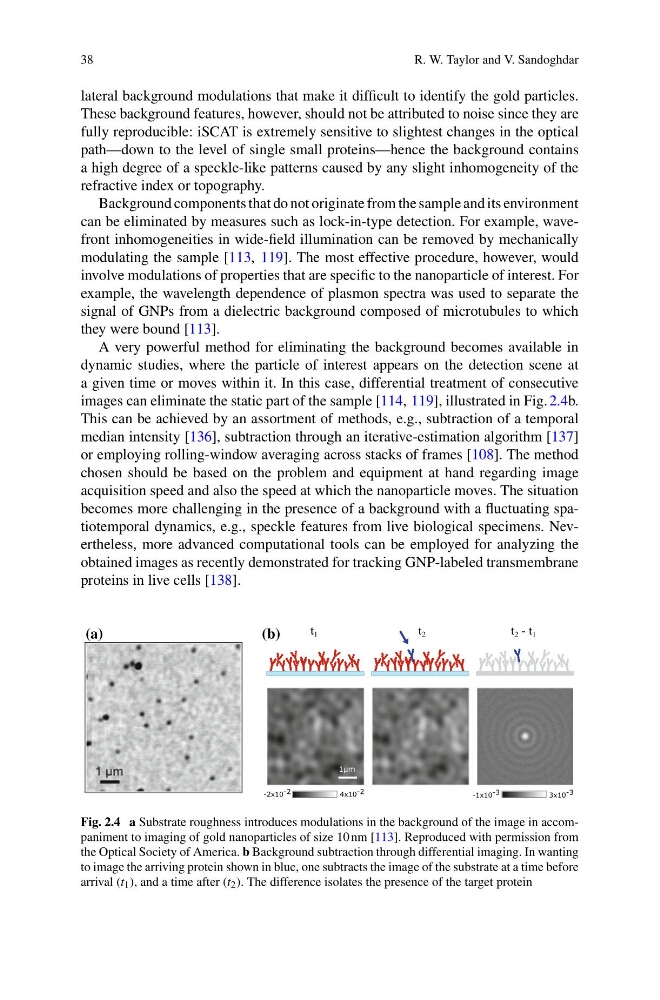
lateral background modulations that make it difficult to identify the gold particles.
These background features, however, should not be attributed to noise since they are
fully reproducible: iSCAT is extremely sensitive to slightest changes in the optical
path—down to the level of single small proteins—hence the background contains
a high degree of a speckle-like patterns caused by any slight inhomogeneity of the
refractive index or topography.
Background components that do not originate from the sample and its environment
can be eliminated by measures such as lock-in-type detection. For example, wavefront inhomogeneities in wide-field illumination can be removed by mechanically
modulating the sample [113, 119]. The most effective procedure, however, would
involve modulations of properties that are specific to the nanoparticle of interest. For
example, the wavelength dependence of plasmon spectra was used to separate the
signal of GNPs from a dielectric background composed of microtubules to which
they were bound [113].
A very powerful method for eliminating the background becomes available in
dynamic studies, where the particle of interest appears on the detection scene at
a given time or moves within it. In this case, differential treatment of consecutive
images can eliminate the static part of the sample [114, 119], illustrated in Fig. 2.4b.
This can be achieved by an assortment of methods, e.g., subtraction of a temporal
median intensity [136], subtraction through an iterative-estimation algorithm [137]
or employing rolling-window averaging across stacks of frames [108]. The method
chosen should be based on the problem and equipment at hand regarding image
acquisition speed and also the speed at which the nanoparticle moves. The situation
becomes more challenging in the presence of a background with a fluctuating spatiotemporal dynamics, e.g., speckle features from live biological specimens. Nevertheless, more advanced computational tools can be employed for analyzing the
obtained images as recently demonstrated for tracking GNP-labeled transmembrane
proteins in live cells [138].
(b)
(a)
t 1
t 2
t 2 - t 1
-2x10 -2
4x10 -2
-1x10 -3
3x10 -3
1μm
Fig. 2.4 a Substrate roughness introduces modulations in the background of the image in accompaniment to imaging of gold nanoparticles of size 10 nm [113]. Reproduced with permission from
the Optical Society of America. b Background subtraction through differential imaging. In wanting
to image the arriving protein shown in blue, one subtracts the image of the substrate at a time before
arrival (t 1 ), and a time after (t 2 ). The difference isolates the presence of the target protein
R. W. Taylor and V. Sandoghdar
lateral background modulations that make it difficult to identify the gold particles.
These background features, however, should not be attributed to noise since they are
fully reproducible: iSCAT is extremely sensitive to slightest changes in the optical
path—down to the level of single small proteins—hence the background contains
a high degree of a speckle-like patterns caused by any slight inhomogeneity of the
refractive index or topography.
Background components that do not originate from the sample and its environment
can be eliminated by measures such as lock-in-type detection. For example, wavefront inhomogeneities in wide-field illumination can be removed by mechanically
modulating the sample [113, 119]. The most effective procedure, however, would
involve modulations of properties that are specific to the nanoparticle of interest. For
example, the wavelength dependence of plasmon spectra was used to separate the
signal of GNPs from a dielectric background composed of microtubules to which
they were bound [113].
A very powerful method for eliminating the background becomes available in
dynamic studies, where the particle of interest appears on the detection scene at
a given time or moves within it. In this case, differential treatment of consecutive
images can eliminate the static part of the sample [114, 119], illustrated in Fig. 2.4b.
This can be achieved by an assortment of methods, e.g., subtraction of a temporal
median intensity [136], subtraction through an iterative-estimation algorithm [137]
or employing rolling-window averaging across stacks of frames [108]. The method
chosen should be based on the problem and equipment at hand regarding image
acquisition speed and also the speed at which the nanoparticle moves. The situation
becomes more challenging in the presence of a background with a fluctuating spatiotemporal dynamics, e.g., speckle features from live biological specimens. Nevertheless, more advanced computational tools can be employed for analyzing the
obtained images as recently demonstrated for tracking GNP-labeled transmembrane
proteins in live cells [138].
(b)
(a)
t 1
t 2
t 2 - t 1
-2x10 -2
4x10 -2
-1x10 -3
3x10 -3
1μm
Fig. 2.4 a Substrate roughness introduces modulations in the background of the image in accompaniment to imaging of gold nanoparticles of size 10 nm [113]. Reproduced with permission from
the Optical Society of America. b Background subtraction through differential imaging. In wanting
to image the arriving protein shown in blue, one subtracts the image of the substrate at a time before
arrival (t 1 ), and a time after (t 2 ). The difference isolates the presence of the target protein