

17 Microsphere-Assisted Interference Microscopy
457
due to their exponential irradiance decrease and, thus, the loss in the high-frequency
information from the sample.
17.2.4 Applications
Full-field microsphere-assisted microscopy has successfully been demonstrated in
several imaging approaches in both the transmission and reflexion modes [134]
ranging from fluorescence nanoscopy [135] to dark-field nanoscopy [136, 137].
Moreover, the imaging technique was applied in various domains such as biological imaging for the visualization of proteins [117], subcellular structures [135],
and medium-sized nonenveloped viruses [138]. After sub-diffraction-limited characterization of materials was first implemented in 2D using microspheres, the technique was then extended to 3D object inspection. This was achieved by combining
a microsphere with interference microscopy in order to measure and reconstruct
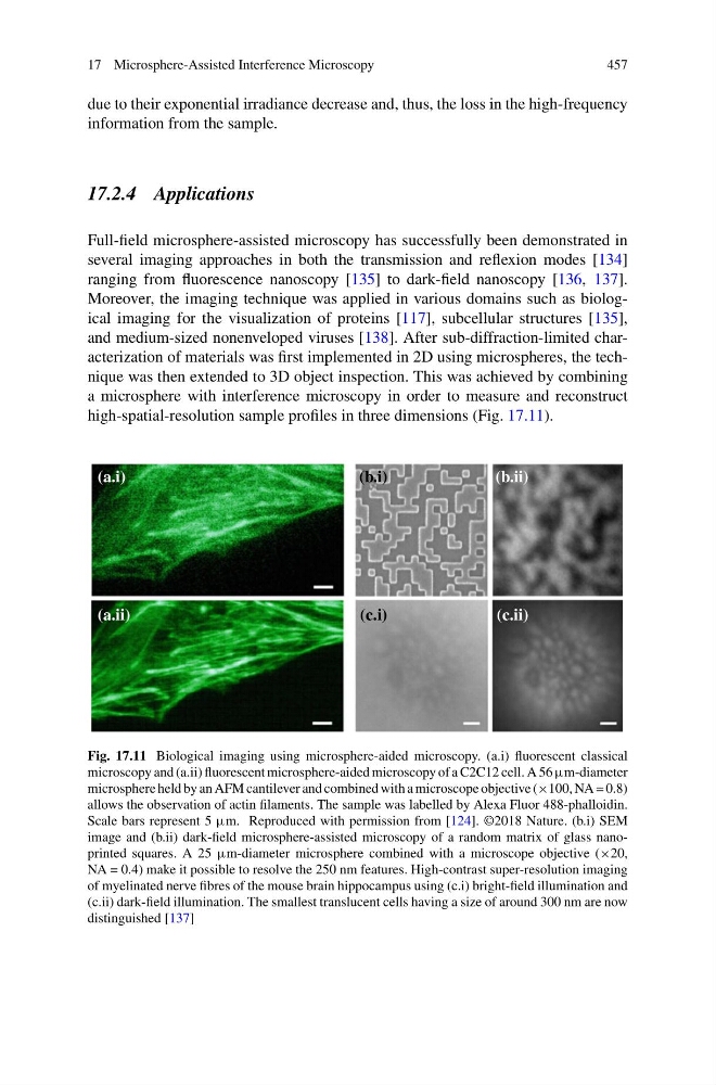
high-spatial-resolution sample profiles in three dimensions (Fig. 17.11).
(a.i)
(b.i)
(a.ii)
(c.i)
(b.ii)
(c.ii)
Fig. 17.11 Biological imaging using microsphere-aided microscopy. (a.i) fluorescent classical
microscopy and (a.ii) fluorescent microsphere-aided microscopy of a C2C12 cell. A 56 µm-diameter
microsphere held by an AFM cantilever and combined with a microscope objective (×100, NA = 0.8)
allows the observation of actin filaments. The sample was labelled by Alexa Fluor 488-phalloidin.
Scale bars represent 5 µm. Reproduced with permission from [124]. ©2018 Nature. (b.i) SEM
image and (b.ii) dark-field microsphere-assisted microscopy of a random matrix of glass nanoprinted squares. A 25 µm-diameter microsphere combined with a microscope objective (×20,
NA = 0.4) make it possible to resolve the 250 nm features. High-contrast super-resolution imaging
of myelinated nerve fibres of the mouse brain hippocampus using (c.i) bright-field illumination and
(c.ii) dark-field illumination. The smallest translucent cells having a size of around 300 nm are now
distinguished [137]
457
due to their exponential irradiance decrease and, thus, the loss in the high-frequency
information from the sample.
17.2.4 Applications
Full-field microsphere-assisted microscopy has successfully been demonstrated in
several imaging approaches in both the transmission and reflexion modes [134]
ranging from fluorescence nanoscopy [135] to dark-field nanoscopy [136, 137].
Moreover, the imaging technique was applied in various domains such as biological imaging for the visualization of proteins [117], subcellular structures [135],
and medium-sized nonenveloped viruses [138]. After sub-diffraction-limited characterization of materials was first implemented in 2D using microspheres, the technique was then extended to 3D object inspection. This was achieved by combining
a microsphere with interference microscopy in order to measure and reconstruct
high-spatial-resolution sample profiles in three dimensions (Fig. 17.11).
(a.i)
(b.i)
(a.ii)
(c.i)
(b.ii)
(c.ii)
Fig. 17.11 Biological imaging using microsphere-aided microscopy. (a.i) fluorescent classical
microscopy and (a.ii) fluorescent microsphere-aided microscopy of a C2C12 cell. A 56 µm-diameter
microsphere held by an AFM cantilever and combined with a microscope objective (×100, NA = 0.8)
allows the observation of actin filaments. The sample was labelled by Alexa Fluor 488-phalloidin.
Scale bars represent 5 µm. Reproduced with permission from [124]. ©2018 Nature. (b.i) SEM
image and (b.ii) dark-field microsphere-assisted microscopy of a random matrix of glass nanoprinted squares. A 25 µm-diameter microsphere combined with a microscope objective (×20,
NA = 0.4) make it possible to resolve the 250 nm features. High-contrast super-resolution imaging
of myelinated nerve fibres of the mouse brain hippocampus using (c.i) bright-field illumination and
(c.ii) dark-field illumination. The smallest translucent cells having a size of around 300 nm are now
distinguished [137]