

16 Theoretical Foundations of Super-Resolution in Microspherical Nanoscopy
411
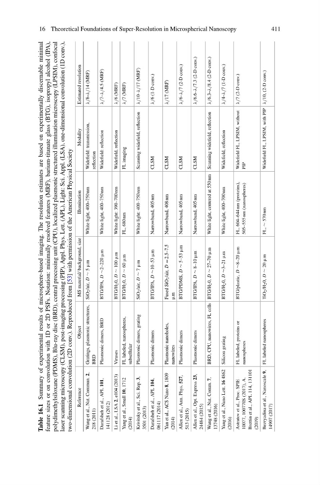
Table 16.1
Summary of experimental results of microsphere-based imaging. The resolution estimates are based on experimentally discernable minimal
feature sizes or on convolution with 1D or 2D PSFs. Notation: minimally resolved features (MRF), barium-titanate glass (BTG), isopropyl alcohol (IPA),
polydimethylsiloxane (PDMS), Blu-ray disc (BRD), central processing unit (CPU), localized plasmonic structured illumination microscopy (LPSIM), confocal
laser scanning microscopy (CLSM), post-imaging processing (PIP), Appl. Phys. Lett. (APL), Light: Sci. Appl. (LSA), one-dimensional convolution (1D conv.),
two-dimensional convolution (2D conv.). Reproduced from [53] with permission of the American Physical Society
Reference
Object
MS material/ background, size
Illumination
Modality
Estimated resolution
Wang et al., Nat. Commun. 2,
218 (2011)
Gratings, plasmonic structures,
BRD
SiO2/air, D
∼ 5
µm
White light, 400–750 nm
Widefield: transmission,
reflection
λ/8–λ/14 (MRF)
Darafsheh et al., APL 101,
141128 (2012)
Plasmonic dimers, BRD
BTG/IPA, D
∼2–220 µm
White light, 400–750 nm
Widefield: reflection
λ/7–λ/4.5 (MRF)
Li et al., LSA 2,
e104 (2013)
Viruses
BTG/H20, D
∼ 100
µm
White light: 390–700 nm
Widefield, reflection
λ/6 (MRF)
Yang et al., Small 10,
1712
(2014)
FL labeled, nanospheres,
subsellular
BTG/H20, D
∼ 60
µm
FL, 680 nm
FL imaging
λ/7 (MRF)
Krivitsky et al., Sci. Rep. 3,
3501 (2013)
Plasmonic dimers, grating
SiO2/air, D
∼ 7
µm
White light: 400–750 nm
Scanning widefield, reflection
λ/10–λ/17 (MRF)
Darafsheh et al., APL 104,
061117 (2014)
Plasmonic dimers
BTG/IPA, D
∼10–53 µm
Narrowband, 405 nm
CLSM
λ/6 (1-D conv.)
Yan et al., ACS Nano 8,
1809
(2014)
Plasmonic nanoholes,
nanowires
Fused SiO2/air, D
= 2.5–7.5
µm
Narrowband, 408 nm
CLSM
λ/17 (MRF)
Allen et al., Ann. Phys. 527,
513 (2015)
Plasmonic dimers
BTG/PDMS, D
∼ 5–53
µm
Narrowband, 405 nm
CLSM
λ/6–λ/7 (2-D conv.)
Allen et al., Opt. Express 23,
24484 (2015)
Plasmonic dimers
BTG/IPA, D
∼ 8–10
µm
Narrowband, 405 nm
CLSM
λ/6.6–λ/7.3 (2-D conv.)
Wang et al., Nat. Comm. 7,
13748 (2016)
BRD, CPU, nanowires, FL cells BTG/H20, D
∼ 27–70
µm
White light, centered at 550 nm Scanning widefield, reflection
λ/6.3–λ/8.4 (2-D conv.)
Yang et al., Nano Lett. 16
4862
(2016)
Silicon grating
BTG/H20, D
∼3–21 µm
White light, 400–700 nm
Widefield, reflection
λ/4–λ/7 (1-D conv.)
Astratov et al., Proc. SPIE
10077, 100770S (2017), A.
Brettin et al., APL 114, 131101
(2019)
FL labeled, proteins or
nanospheres
BTG/plastic, D
∼8–20 µm
FL, 604–644 nm (proteins),
505–555 nm (nanospheres)
Widefield FL, LPSIM, without
PIP
λ/7 (2-D conv.)
Bezryadina et al., Nanoscale 9,
14907 (2017)
FL labeled nanospheres
TiO2/H20, D
∼ 20
µm
FL,
∼ 570 nm
Widefield FL, LPSIM, with PIP
λ/10, (2-D conv.)
411
Table 16.1
Summary of experimental results of microsphere-based imaging. The resolution estimates are based on experimentally discernable minimal
feature sizes or on convolution with 1D or 2D PSFs. Notation: minimally resolved features (MRF), barium-titanate glass (BTG), isopropyl alcohol (IPA),
polydimethylsiloxane (PDMS), Blu-ray disc (BRD), central processing unit (CPU), localized plasmonic structured illumination microscopy (LPSIM), confocal
laser scanning microscopy (CLSM), post-imaging processing (PIP), Appl. Phys. Lett. (APL), Light: Sci. Appl. (LSA), one-dimensional convolution (1D conv.),
two-dimensional convolution (2D conv.). Reproduced from [53] with permission of the American Physical Society
Reference
Object
MS material/ background, size
Illumination
Modality
Estimated resolution
Wang et al., Nat. Commun. 2,
218 (2011)
Gratings, plasmonic structures,
BRD
SiO2/air, D
∼ 5
µm
White light, 400–750 nm
Widefield: transmission,
reflection
λ/8–λ/14 (MRF)
Darafsheh et al., APL 101,
141128 (2012)
Plasmonic dimers, BRD
BTG/IPA, D
∼2–220 µm
White light, 400–750 nm
Widefield: reflection
λ/7–λ/4.5 (MRF)
Li et al., LSA 2,
e104 (2013)
Viruses
BTG/H20, D
∼ 100
µm
White light: 390–700 nm
Widefield, reflection
λ/6 (MRF)
Yang et al., Small 10,
1712
(2014)
FL labeled, nanospheres,
subsellular
BTG/H20, D
∼ 60
µm
FL, 680 nm
FL imaging
λ/7 (MRF)
Krivitsky et al., Sci. Rep. 3,
3501 (2013)
Plasmonic dimers, grating
SiO2/air, D
∼ 7
µm
White light: 400–750 nm
Scanning widefield, reflection
λ/10–λ/17 (MRF)
Darafsheh et al., APL 104,
061117 (2014)
Plasmonic dimers
BTG/IPA, D
∼10–53 µm
Narrowband, 405 nm
CLSM
λ/6 (1-D conv.)
Yan et al., ACS Nano 8,
1809
(2014)
Plasmonic nanoholes,
nanowires
Fused SiO2/air, D
= 2.5–7.5
µm
Narrowband, 408 nm
CLSM
λ/17 (MRF)
Allen et al., Ann. Phys. 527,
513 (2015)
Plasmonic dimers
BTG/PDMS, D
∼ 5–53
µm
Narrowband, 405 nm
CLSM
λ/6–λ/7 (2-D conv.)
Allen et al., Opt. Express 23,
24484 (2015)
Plasmonic dimers
BTG/IPA, D
∼ 8–10
µm
Narrowband, 405 nm
CLSM
λ/6.6–λ/7.3 (2-D conv.)
Wang et al., Nat. Comm. 7,
13748 (2016)
BRD, CPU, nanowires, FL cells BTG/H20, D
∼ 27–70
µm
White light, centered at 550 nm Scanning widefield, reflection
λ/6.3–λ/8.4 (2-D conv.)
Yang et al., Nano Lett. 16
4862
(2016)
Silicon grating
BTG/H20, D
∼3–21 µm
White light, 400–700 nm
Widefield, reflection
λ/4–λ/7 (1-D conv.)
Astratov et al., Proc. SPIE
10077, 100770S (2017), A.
Brettin et al., APL 114, 131101
(2019)
FL labeled, proteins or
nanospheres
BTG/plastic, D
∼8–20 µm
FL, 604–644 nm (proteins),
505–555 nm (nanospheres)
Widefield FL, LPSIM, without
PIP
λ/7 (2-D conv.)
Bezryadina et al., Nanoscale 9,
14907 (2017)
FL labeled nanospheres
TiO2/H20, D
∼ 20
µm
FL,
∼ 570 nm
Widefield FL, LPSIM, with PIP
λ/10, (2-D conv.)