

374
Z. Wang and B. Luk’yanchuk
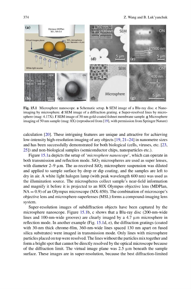
Fig. 15.1 Microsphere nanoscope. a Schematic setup. b SEM image of a Blu-ray disc. c Nanoimaging by microsphere. d SEM image of a diffraction grating. e Super-resolved lines by microsphere (mag: 4.17X). f SEM image of 50 nm gold-coated fishnet membrane sample. g Microsphere
imaging of 50 nm sample (mag: 8X) (reproduced from [19], with permission from Springer Nature)
calculation [20]. These intriguing features are unique and attractive for achieving
low-intensity high-resolution imaging of any objects [19, 21–24] in nanometre sizes
and has been successfully demonstrated for both biological (cells, viruses, etc. [23,
25]) and non-biological samples (semiconductor chips, nanoparticles etc.).
Figure 15.1a depicts the setup of ‘microsphere nanoscope’, which can operate in
both transmission and reflection mode. SiO 2 microspheres are used as super lenses,
with diameter 2–9 μm. The as-received SiO 2 microsphere suspension was diluted
and applied to sample surface by drop or dip coating, and the samples are left to
dry in air. A white light halogen lamp (with peak wavelength 600 nm) was used as
the illumination source. The microspheres collect sample’s near-field information
and magnify it before it is projected to an 80X Olympus objective lens (MDPlan,
NA = 0.9) of an Olympus microscope (MX-850). The combination of microscope’s
objective lens and microsphere-superlenses (MSL) forms a compound-imaging lens
system.
Super-resolution images of subdiffraction objects have been captured by the
microsphere nanoscope. Figure 15.1b, c shows that a Blu-ray disc (200-nm-wide
lines and 100-nm-wide grooves) are clearly imaged by a 4.7 μm microsphere in
reflection mode. In another example (Fig. 15.1d, e), the diffraction gratings (coated
with 30-nm thick chrome-film, 360-nm-wide lines spaced 130 nm apart on fused
silica substrates) were imaged in transmission mode. Only lines with microsphere
particles placed on top were resolved. The lines without the particles mix together and
form a bright spot that cannot be directly resolved by the optical microscope because
of the diffraction limit. The virtual image plane was 2.5 μm beneath the sample
surface. These images are in super-resolution, because the best diffraction-limited
Z. Wang and B. Luk’yanchuk
Fig. 15.1 Microsphere nanoscope. a Schematic setup. b SEM image of a Blu-ray disc. c Nanoimaging by microsphere. d SEM image of a diffraction grating. e Super-resolved lines by microsphere (mag: 4.17X). f SEM image of 50 nm gold-coated fishnet membrane sample. g Microsphere
imaging of 50 nm sample (mag: 8X) (reproduced from [19], with permission from Springer Nature)
calculation [20]. These intriguing features are unique and attractive for achieving
low-intensity high-resolution imaging of any objects [19, 21–24] in nanometre sizes
and has been successfully demonstrated for both biological (cells, viruses, etc. [23,
25]) and non-biological samples (semiconductor chips, nanoparticles etc.).
Figure 15.1a depicts the setup of ‘microsphere nanoscope’, which can operate in
both transmission and reflection mode. SiO 2 microspheres are used as super lenses,
with diameter 2–9 μm. The as-received SiO 2 microsphere suspension was diluted
and applied to sample surface by drop or dip coating, and the samples are left to
dry in air. A white light halogen lamp (with peak wavelength 600 nm) was used as
the illumination source. The microspheres collect sample’s near-field information
and magnify it before it is projected to an 80X Olympus objective lens (MDPlan,
NA = 0.9) of an Olympus microscope (MX-850). The combination of microscope’s
objective lens and microsphere-superlenses (MSL) forms a compound-imaging lens
system.
Super-resolution images of subdiffraction objects have been captured by the
microsphere nanoscope. Figure 15.1b, c shows that a Blu-ray disc (200-nm-wide
lines and 100-nm-wide grooves) are clearly imaged by a 4.7 μm microsphere in
reflection mode. In another example (Fig. 15.1d, e), the diffraction gratings (coated
with 30-nm thick chrome-film, 360-nm-wide lines spaced 130 nm apart on fused
silica substrates) were imaged in transmission mode. Only lines with microsphere
particles placed on top were resolved. The lines without the particles mix together and
form a bright spot that cannot be directly resolved by the optical microscope because
of the diffraction limit. The virtual image plane was 2.5 μm beneath the sample
surface. These images are in super-resolution, because the best diffraction-limited