

12 Nonlinear Label-Free Super-Resolution Microscopy Using Structured Illumination
305
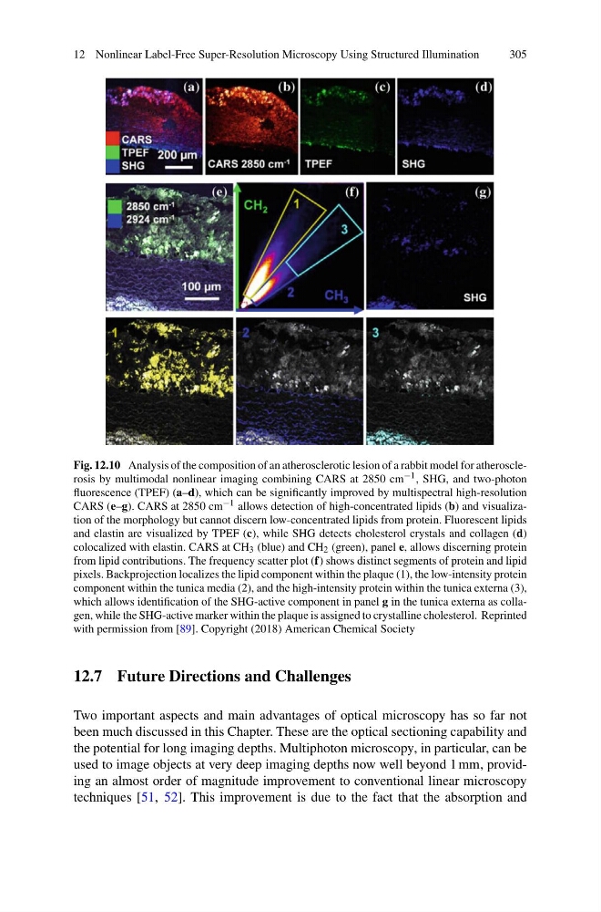
Fig. 12.10 Analysis of the composition of an atherosclerotic lesion of a rabbit model for atherosclerosis by multimodal nonlinear imaging combining CARS at 2850 cm −1 , SHG, and two-photon
fluorescence (TPEF) (a–d), which can be significantly improved by multispectral high-resolution
CARS (e–g). CARS at 2850 cm −1 allows detection of high-concentrated lipids (b) and visualization of the morphology but cannot discern low-concentrated lipids from protein. Fluorescent lipids
and elastin are visualized by TPEF (c), while SHG detects cholesterol crystals and collagen (d)
colocalized with elastin. CARS at CH 3 (blue) and CH 2 (green), panel e, allows discerning protein
from lipid contributions. The frequency scatter plot (f) shows distinct segments of protein and lipid
pixels. Backprojection localizes the lipid component within the plaque (1), the low-intensity protein
component within the tunica media (2), and the high-intensity protein within the tunica externa (3),
which allows identification of the SHG-active component in panel g in the tunica externa as collagen, while the SHG-active marker within the plaque is assigned to crystalline cholesterol. Reprinted
with permission from [89]. Copyright (2018) American Chemical Society
12.7 Future Directions and Challenges
Two important aspects and main advantages of optical microscopy has so far not
been much discussed in this Chapter. These are the optical sectioning capability and
the potential for long imaging depths. Multiphoton microscopy, in particular, can be
used to image objects at very deep imaging depths now well beyond 1 mm, providing an almost order of magnitude improvement to conventional linear microscopy
techniques [51, 52]. This improvement is due to the fact that the absorption and
305
Fig. 12.10 Analysis of the composition of an atherosclerotic lesion of a rabbit model for atherosclerosis by multimodal nonlinear imaging combining CARS at 2850 cm −1 , SHG, and two-photon
fluorescence (TPEF) (a–d), which can be significantly improved by multispectral high-resolution
CARS (e–g). CARS at 2850 cm −1 allows detection of high-concentrated lipids (b) and visualization of the morphology but cannot discern low-concentrated lipids from protein. Fluorescent lipids
and elastin are visualized by TPEF (c), while SHG detects cholesterol crystals and collagen (d)
colocalized with elastin. CARS at CH 3 (blue) and CH 2 (green), panel e, allows discerning protein
from lipid contributions. The frequency scatter plot (f) shows distinct segments of protein and lipid
pixels. Backprojection localizes the lipid component within the plaque (1), the low-intensity protein
component within the tunica media (2), and the high-intensity protein within the tunica externa (3),
which allows identification of the SHG-active component in panel g in the tunica externa as collagen, while the SHG-active marker within the plaque is assigned to crystalline cholesterol. Reprinted
with permission from [89]. Copyright (2018) American Chemical Society
12.7 Future Directions and Challenges
Two important aspects and main advantages of optical microscopy has so far not
been much discussed in this Chapter. These are the optical sectioning capability and
the potential for long imaging depths. Multiphoton microscopy, in particular, can be
used to image objects at very deep imaging depths now well beyond 1 mm, providing an almost order of magnitude improvement to conventional linear microscopy
techniques [51, 52]. This improvement is due to the fact that the absorption and