

178
P. Bianchini et al.
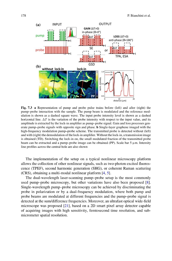
Fig. 7.3 a Representation of pump and probe pulse trains before (left) and after (right) the
pump–probe interaction with the sample. The pump beam is modulated and the reference modulation is shown as a dashed square wave. The input probe intensity level is shown as a dashed
horizontal line. T is the variation of the probe intensity with respect to the input value, and its
amplitude is extracted by the lock-in amplifier as pump–probe signal. Gain and loss processes generate pump–probe signals with opposite sign and phase. b Single-layer graphene imaged with the
high-frequency modulation pump–probe scheme. The transmitted probe is detected without (left)
and with (right) the demodulation of the lock-in amplifier. Without the lock-in, a transmission image
is obtained (TD). Switching the lock-in on, the small modulated fraction of the transmitted probe
beam can be extracted and a pump–probe image can be obtained (PP). Scale bar 5 μm. Intensity
line profiles across the central hole are also shown
The implementation of the setup on a typical nonlinear microscopy platform
allows the collection of other nonlinear signals, such as two-photon excited fluorescence (TPEF), second harmonic generation (SHG), or coherent Raman scattering
(CRS), obtaining a multi-modal nonlinear platform [4, 5].
The dual-wavelength laser-scanning pump–probe setup is the most commonly
used pump–probe microscopy, but other variations have also been proposed [8].
Single-wavelength pump–probe microscopy can be achieved by discriminating the
probe in polarization or by a dual-frequency modulation, where both pump and
probe beams are modulated at different frequencies and the pump–probe signal is
detected at the sum/difference frequencies. Moreover, an ultrafast optical wide-field
microscope was proposed [21], based on a 2D smart pixel array detector capable
of acquiring images with high sensitivity, femtosecond time resolution, and submicrometer spatial resolution.
P. Bianchini et al.
Fig. 7.3 a Representation of pump and probe pulse trains before (left) and after (right) the
pump–probe interaction with the sample. The pump beam is modulated and the reference modulation is shown as a dashed square wave. The input probe intensity level is shown as a dashed
horizontal line. T is the variation of the probe intensity with respect to the input value, and its
amplitude is extracted by the lock-in amplifier as pump–probe signal. Gain and loss processes generate pump–probe signals with opposite sign and phase. b Single-layer graphene imaged with the
high-frequency modulation pump–probe scheme. The transmitted probe is detected without (left)
and with (right) the demodulation of the lock-in amplifier. Without the lock-in, a transmission image
is obtained (TD). Switching the lock-in on, the small modulated fraction of the transmitted probe
beam can be extracted and a pump–probe image can be obtained (PP). Scale bar 5 μm. Intensity
line profiles across the central hole are also shown
The implementation of the setup on a typical nonlinear microscopy platform
allows the collection of other nonlinear signals, such as two-photon excited fluorescence (TPEF), second harmonic generation (SHG), or coherent Raman scattering
(CRS), obtaining a multi-modal nonlinear platform [4, 5].
The dual-wavelength laser-scanning pump–probe setup is the most commonly
used pump–probe microscopy, but other variations have also been proposed [8].
Single-wavelength pump–probe microscopy can be achieved by discriminating the
probe in polarization or by a dual-frequency modulation, where both pump and
probe beams are modulated at different frequencies and the pump–probe signal is
detected at the sum/difference frequencies. Moreover, an ultrafast optical wide-field
microscope was proposed [21], based on a 2D smart pixel array detector capable
of acquiring images with high sensitivity, femtosecond time resolution, and submicrometer spatial resolution.