

6 Absorption-Based Far-Field Label-Free Super-Resolution …
153
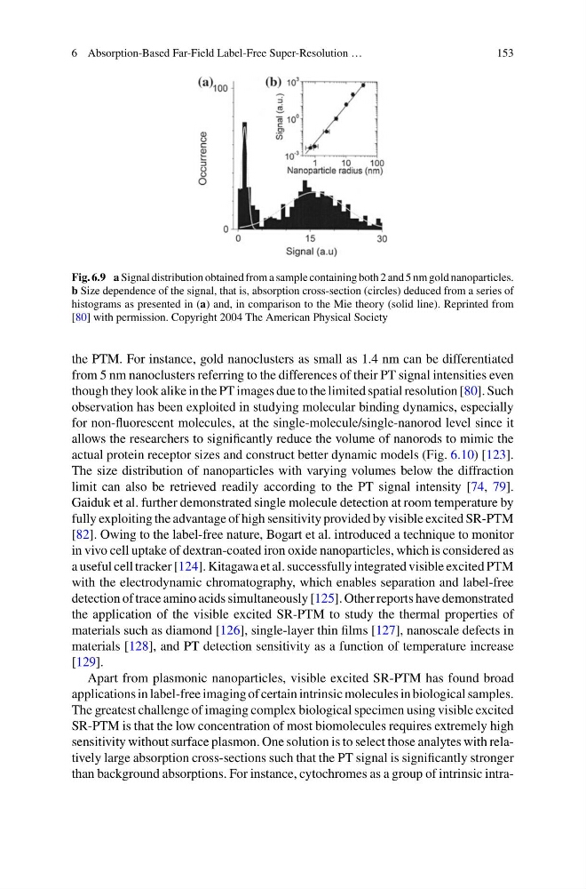
Fig. 6.9 a Signal distribution obtained from a sample containing both 2 and 5 nm gold nanoparticles.
b Size dependence of the signal, that is, absorption cross-section (circles) deduced from a series of
histograms as presented in (a) and, in comparison to the Mie theory (solid line). Reprinted from
[80] with permission. Copyright 2004 The American Physical Society
the PTM. For instance, gold nanoclusters as small as 1.4 nm can be differentiated
from 5 nm nanoclusters referring to the differences of their PT signal intensities even
though they look alike in the PT images due to the limited spatial resolution [80]. Such
observation has been exploited in studying molecular binding dynamics, especially
for non-fluorescent molecules, at the single-molecule/single-nanorod level since it
allows the researchers to significantly reduce the volume of nanorods to mimic the
actual protein receptor sizes and construct better dynamic models (Fig. 6.10) [123].
The size distribution of nanoparticles with varying volumes below the diffraction
limit can also be retrieved readily according to the PT signal intensity [74, 79].
Gaiduk et al. further demonstrated single molecule detection at room temperature by
fully exploiting the advantage of high sensitivity provided by visible excited SR-PTM
[82]. Owing to the label-free nature, Bogart et al. introduced a technique to monitor
in vivo cell uptake of dextran-coated iron oxide nanoparticles, which is considered as
a useful cell tracker [124]. Kitagawa et al. successfully integrated visible excited PTM
with the electrodynamic chromatography, which enables separation and label-free
detection of trace amino acids simultaneously [125]. Other reports have demonstrated
the application of the visible excited SR-PTM to study the thermal properties of
materials such as diamond [126], single-layer thin films [127], nanoscale defects in
materials [128], and PT detection sensitivity as a function of temperature increase
[129].
Apart from plasmonic nanoparticles, visible excited SR-PTM has found broad
applications in label-free imaging of certain intrinsic molecules in biological samples.
The greatest challenge of imaging complex biological specimen using visible excited
SR-PTM is that the low concentration of most biomolecules requires extremely high
sensitivity without surface plasmon. One solution is to select those analytes with relatively large absorption cross-sections such that the PT signal is significantly stronger
than background absorptions. For instance, cytochromes as a group of intrinsic intra-
153
Fig. 6.9 a Signal distribution obtained from a sample containing both 2 and 5 nm gold nanoparticles.
b Size dependence of the signal, that is, absorption cross-section (circles) deduced from a series of
histograms as presented in (a) and, in comparison to the Mie theory (solid line). Reprinted from
[80] with permission. Copyright 2004 The American Physical Society
the PTM. For instance, gold nanoclusters as small as 1.4 nm can be differentiated
from 5 nm nanoclusters referring to the differences of their PT signal intensities even
though they look alike in the PT images due to the limited spatial resolution [80]. Such
observation has been exploited in studying molecular binding dynamics, especially
for non-fluorescent molecules, at the single-molecule/single-nanorod level since it
allows the researchers to significantly reduce the volume of nanorods to mimic the
actual protein receptor sizes and construct better dynamic models (Fig. 6.10) [123].
The size distribution of nanoparticles with varying volumes below the diffraction
limit can also be retrieved readily according to the PT signal intensity [74, 79].
Gaiduk et al. further demonstrated single molecule detection at room temperature by
fully exploiting the advantage of high sensitivity provided by visible excited SR-PTM
[82]. Owing to the label-free nature, Bogart et al. introduced a technique to monitor
in vivo cell uptake of dextran-coated iron oxide nanoparticles, which is considered as
a useful cell tracker [124]. Kitagawa et al. successfully integrated visible excited PTM
with the electrodynamic chromatography, which enables separation and label-free
detection of trace amino acids simultaneously [125]. Other reports have demonstrated
the application of the visible excited SR-PTM to study the thermal properties of
materials such as diamond [126], single-layer thin films [127], nanoscale defects in
materials [128], and PT detection sensitivity as a function of temperature increase
[129].
Apart from plasmonic nanoparticles, visible excited SR-PTM has found broad
applications in label-free imaging of certain intrinsic molecules in biological samples.
The greatest challenge of imaging complex biological specimen using visible excited
SR-PTM is that the low concentration of most biomolecules requires extremely high
sensitivity without surface plasmon. One solution is to select those analytes with relatively large absorption cross-sections such that the PT signal is significantly stronger
than background absorptions. For instance, cytochromes as a group of intrinsic intra-