

80
C.-L. Hsieh
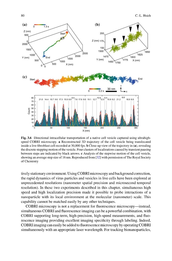
Fig. 3.6 Directional intracellular transportation of a native cell vesicle captured using ultrahighspeed COBRI microscopy. a Reconstructed 3D trajectory of the cell vesicle being translocated
inside a live fibroblast cell recorded at 30,000 fps. b Close-up view of the trajectory in (a), revealing
the discrete stepping motion of the vesicle. Four clusters of localizations caused by transient pausing
between steps are indicated by black arrows. c Analysis of the stepwise motion of the cell vesicle,
showing an average step size of 16 nm. Reproduced from [32] with permission of The Royal Society
of Chemistry
tively stationary environment. Using COBRI microscopy and background correction,
the rapid dynamics of virus particles and vesicles in live cells have been explored at
unprecedented resolutions (nanometer spatial precision and microsecond temporal
resolution). In these two experiments described in this chapter, simultaneous high
speed and high localization precision made it possible to probe interactions of a
nanoparticle with its local environment at the molecular (nanometer) scale. This
capability cannot be matched easily by any other techniques.
COBRI microscopy is not a replacement for fluorescence microscopy—instead,
simultaneous COBRI and fluorescence imaging can be a powerful combination, with
COBRI supporting long-term, high-precision, high-speed measurements, and fluorescence imaging providing excellent imaging specificity through labeling. Indeed,
COBRI imaging can easily be added to fluorescence microscopy by operating COBRI
simultaneously with an appropriate laser wavelength. For tracking bionanoparticles,
C.-L. Hsieh
Fig. 3.6 Directional intracellular transportation of a native cell vesicle captured using ultrahighspeed COBRI microscopy. a Reconstructed 3D trajectory of the cell vesicle being translocated
inside a live fibroblast cell recorded at 30,000 fps. b Close-up view of the trajectory in (a), revealing
the discrete stepping motion of the vesicle. Four clusters of localizations caused by transient pausing
between steps are indicated by black arrows. c Analysis of the stepwise motion of the cell vesicle,
showing an average step size of 16 nm. Reproduced from [32] with permission of The Royal Society
of Chemistry
tively stationary environment. Using COBRI microscopy and background correction,
the rapid dynamics of virus particles and vesicles in live cells have been explored at
unprecedented resolutions (nanometer spatial precision and microsecond temporal
resolution). In these two experiments described in this chapter, simultaneous high
speed and high localization precision made it possible to probe interactions of a
nanoparticle with its local environment at the molecular (nanometer) scale. This
capability cannot be matched easily by any other techniques.
COBRI microscopy is not a replacement for fluorescence microscopy—instead,
simultaneous COBRI and fluorescence imaging can be a powerful combination, with
COBRI supporting long-term, high-precision, high-speed measurements, and fluorescence imaging providing excellent imaging specificity through labeling. Indeed,
COBRI imaging can easily be added to fluorescence microscopy by operating COBRI
simultaneously with an appropriate laser wavelength. For tracking bionanoparticles,