

62
E. Liebenthal and T. Singhal
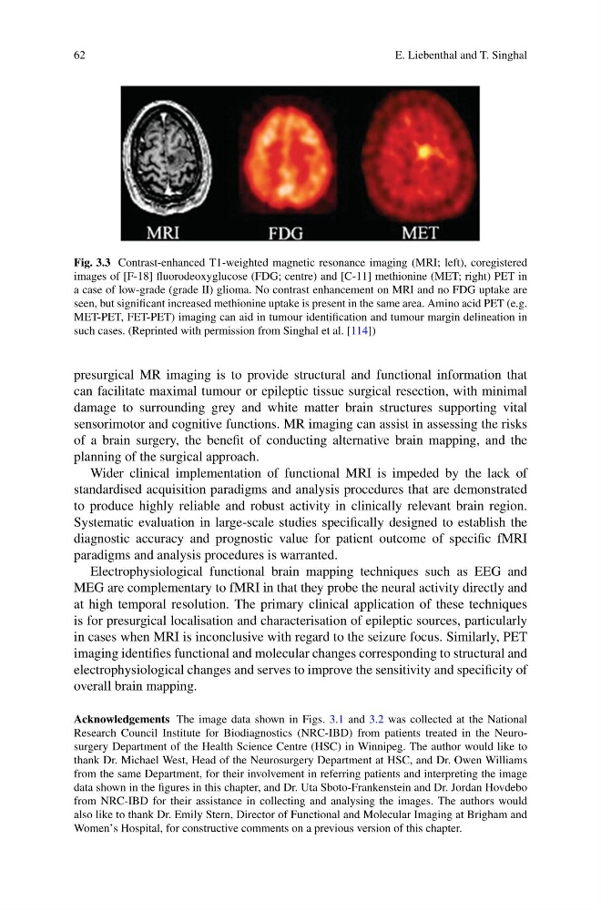
Fig. 3.3 Contrast-enhanced T1-weighted magnetic resonance imaging (MRI; left), coregistered
images of [F-18] fluorodeoxyglucose (FDG; centre) and [C-11] methionine (MET; right) PET in
a case of low-grade (grade II) glioma. No contrast enhancement on MRI and no FDG uptake are
seen, but significant increased methionine uptake is present in the same area. Amino acid PET (e.g.
MET-PET, FET-PET) imaging can aid in tumour identification and tumour margin delineation in
such cases. (Reprinted with permission from Singhal et al. [114])
presurgical MR imaging is to provide structural and functional information that
can facilitate maximal tumour or epileptic tissue surgical resection, with minimal
damage to surrounding grey and white matter brain structures supporting vital
sensorimotor and cognitive functions. MR imaging can assist in assessing the risks
of a brain surgery, the benefit of conducting alternative brain mapping, and the
planning of the surgical approach.
Wider clinical implementation of functional MRI is impeded by the lack of
standardised acquisition paradigms and analysis procedures that are demonstrated
to produce highly reliable and robust activity in clinically relevant brain region.
Systematic evaluation in large-scale studies specifically designed to establish the
diagnostic accuracy and prognostic value for patient outcome of specific fMRI
paradigms and analysis procedures is warranted.
Electrophysiological functional brain mapping techniques such as EEG and
MEG are complementary to fMRI in that they probe the neural activity directly and
at high temporal resolution. The primary clinical application of these techniques
is for presurgical localisation and characterisation of epileptic sources, particularly
in cases when MRI is inconclusive with regard to the seizure focus. Similarly, PET
imaging identifies functional and molecular changes corresponding to structural and
electrophysiological changes and serves to improve the sensitivity and specificity of
overall brain mapping.
Acknowledgements The image data shown in Figs. 3.1 and 3.2 was collected at the National
Research Council Institute for Biodiagnostics (NRC-IBD) from patients treated in the Neurosurgery Department of the Health Science Centre (HSC) in Winnipeg. The author would like to
thank Dr. Michael West, Head of the Neurosurgery Department at HSC, and Dr. Owen Williams
from the same Department, for their involvement in referring patients and interpreting the image
data shown in the figures in this chapter, and Dr. Uta Sboto-Frankenstein and Dr. Jordan Hovdebo
from NRC-IBD for their assistance in collecting and analysing the images. The authors would
also like to thank Dr. Emily Stern, Director of Functional and Molecular Imaging at Brigham and
Women’s Hospital, for constructive comments on a previous version of this chapter.
E. Liebenthal and T. Singhal
Fig. 3.3 Contrast-enhanced T1-weighted magnetic resonance imaging (MRI; left), coregistered
images of [F-18] fluorodeoxyglucose (FDG; centre) and [C-11] methionine (MET; right) PET in
a case of low-grade (grade II) glioma. No contrast enhancement on MRI and no FDG uptake are
seen, but significant increased methionine uptake is present in the same area. Amino acid PET (e.g.
MET-PET, FET-PET) imaging can aid in tumour identification and tumour margin delineation in
such cases. (Reprinted with permission from Singhal et al. [114])
presurgical MR imaging is to provide structural and functional information that
can facilitate maximal tumour or epileptic tissue surgical resection, with minimal
damage to surrounding grey and white matter brain structures supporting vital
sensorimotor and cognitive functions. MR imaging can assist in assessing the risks
of a brain surgery, the benefit of conducting alternative brain mapping, and the
planning of the surgical approach.
Wider clinical implementation of functional MRI is impeded by the lack of
standardised acquisition paradigms and analysis procedures that are demonstrated
to produce highly reliable and robust activity in clinically relevant brain region.
Systematic evaluation in large-scale studies specifically designed to establish the
diagnostic accuracy and prognostic value for patient outcome of specific fMRI
paradigms and analysis procedures is warranted.
Electrophysiological functional brain mapping techniques such as EEG and
MEG are complementary to fMRI in that they probe the neural activity directly and
at high temporal resolution. The primary clinical application of these techniques
is for presurgical localisation and characterisation of epileptic sources, particularly
in cases when MRI is inconclusive with regard to the seizure focus. Similarly, PET
imaging identifies functional and molecular changes corresponding to structural and
electrophysiological changes and serves to improve the sensitivity and specificity of
overall brain mapping.
Acknowledgements The image data shown in Figs. 3.1 and 3.2 was collected at the National
Research Council Institute for Biodiagnostics (NRC-IBD) from patients treated in the Neurosurgery Department of the Health Science Centre (HSC) in Winnipeg. The author would like to
thank Dr. Michael West, Head of the Neurosurgery Department at HSC, and Dr. Owen Williams
from the same Department, for their involvement in referring patients and interpreting the image
data shown in the figures in this chapter, and Dr. Uta Sboto-Frankenstein and Dr. Jordan Hovdebo
from NRC-IBD for their assistance in collecting and analysing the images. The authors would
also like to thank Dr. Emily Stern, Director of Functional and Molecular Imaging at Brigham and
Women’s Hospital, for constructive comments on a previous version of this chapter.