

58
E. Liebenthal and T. Singhal
Right
Left
56L
44L
32L
20L
8L
56R
44R
32R
20R
t
2 4
6
10
8R
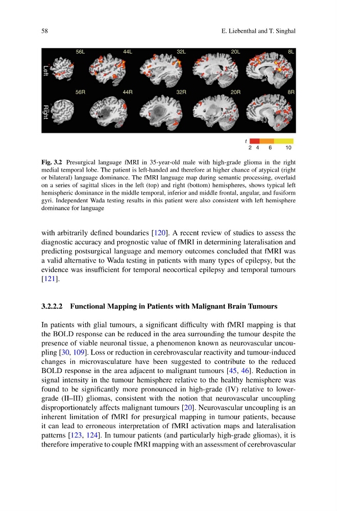
Fig. 3.2 Presurgical language fMRI in 35-year-old male with high-grade glioma in the right
medial temporal lobe. The patient is left-handed and therefore at higher chance of atypical (right
or bilateral) language dominance. The fMRI language map during semantic processing, overlaid
on a series of sagittal slices in the left (top) and right (bottom) hemispheres, shows typical left
hemispheric dominance in the middle temporal, inferior and middle frontal, angular, and fusiform
gyri. Independent Wada testing results in this patient were also consistent with left hemisphere
dominance for language
with arbitrarily defined boundaries [120]. A recent review of studies to assess the
diagnostic accuracy and prognostic value of fMRI in determining lateralisation and
predicting postsurgical language and memory outcomes concluded that fMRI was
a valid alternative to Wada testing in patients with many types of epilepsy, but the
evidence was insufficient for temporal neocortical epilepsy and temporal tumours
[121].
3.2.2.2 Functional Mapping in Patients with Malignant Brain Tumours
In patients with glial tumours, a significant difficulty with fMRI mapping is that
the BOLD response can be reduced in the area surrounding the tumour despite the
presence of viable neuronal tissue, a phenomenon known as neurovascular uncoupling [30, 109]. Loss or reduction in cerebrovascular reactivity and tumour-induced
changes in microvasculature have been suggested to contribute to the reduced
BOLD response in the area adjacent to malignant tumours [45, 46]. Reduction in
signal intensity in the tumour hemisphere relative to the healthy hemisphere was
found to be significantly more pronounced in high-grade (IV) relative to lowergrade (II–III) gliomas, consistent with the notion that neurovascular uncoupling
disproportionately affects malignant tumours [20]. Neurovascular uncoupling is an
inherent limitation of fMRI for presurgical mapping in tumour patients, because
it can lead to erroneous interpretation of fMRI activation maps and lateralisation
patterns [123, 124]. In tumour patients (and particularly high-grade gliomas), it is
therefore imperative to couple fMRI mapping with an assessment of cerebrovascular
E. Liebenthal and T. Singhal
Right
Left
56L
44L
32L
20L
8L
56R
44R
32R
20R
t
2 4
6
10
8R
Fig. 3.2 Presurgical language fMRI in 35-year-old male with high-grade glioma in the right
medial temporal lobe. The patient is left-handed and therefore at higher chance of atypical (right
or bilateral) language dominance. The fMRI language map during semantic processing, overlaid
on a series of sagittal slices in the left (top) and right (bottom) hemispheres, shows typical left
hemispheric dominance in the middle temporal, inferior and middle frontal, angular, and fusiform
gyri. Independent Wada testing results in this patient were also consistent with left hemisphere
dominance for language
with arbitrarily defined boundaries [120]. A recent review of studies to assess the
diagnostic accuracy and prognostic value of fMRI in determining lateralisation and
predicting postsurgical language and memory outcomes concluded that fMRI was
a valid alternative to Wada testing in patients with many types of epilepsy, but the
evidence was insufficient for temporal neocortical epilepsy and temporal tumours
[121].
3.2.2.2 Functional Mapping in Patients with Malignant Brain Tumours
In patients with glial tumours, a significant difficulty with fMRI mapping is that
the BOLD response can be reduced in the area surrounding the tumour despite the
presence of viable neuronal tissue, a phenomenon known as neurovascular uncoupling [30, 109]. Loss or reduction in cerebrovascular reactivity and tumour-induced
changes in microvasculature have been suggested to contribute to the reduced
BOLD response in the area adjacent to malignant tumours [45, 46]. Reduction in
signal intensity in the tumour hemisphere relative to the healthy hemisphere was
found to be significantly more pronounced in high-grade (IV) relative to lowergrade (II–III) gliomas, consistent with the notion that neurovascular uncoupling
disproportionately affects malignant tumours [20]. Neurovascular uncoupling is an
inherent limitation of fMRI for presurgical mapping in tumour patients, because
it can lead to erroneous interpretation of fMRI activation maps and lateralisation
patterns [123, 124]. In tumour patients (and particularly high-grade gliomas), it is
therefore imperative to couple fMRI mapping with an assessment of cerebrovascular