

56
E. Liebenthal and T. Singhal
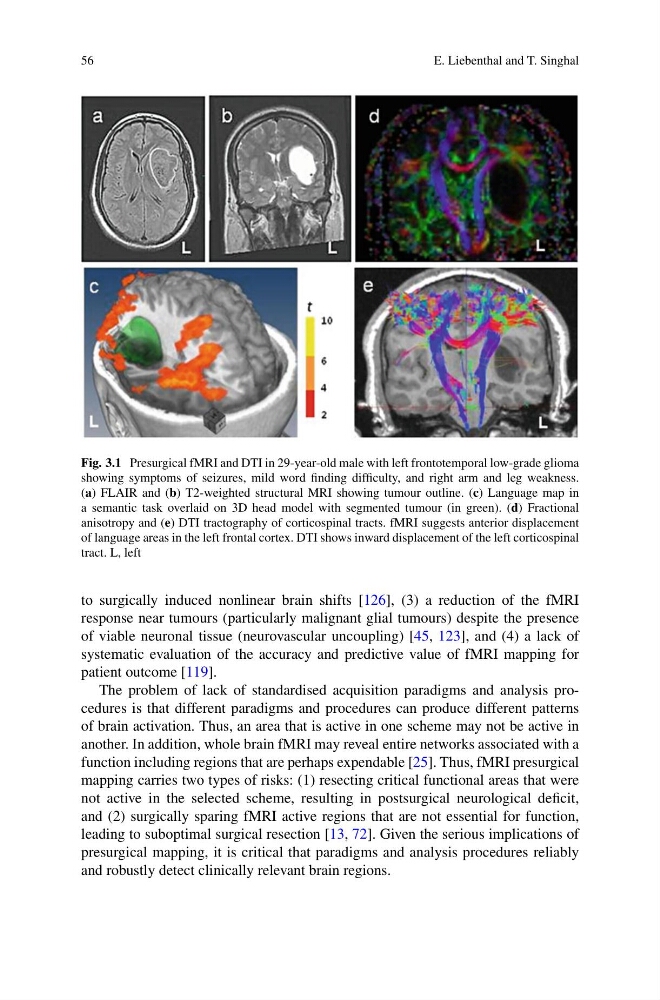
Fig. 3.1 Presurgical fMRI and DTI in 29-year-old male with left frontotemporal low-grade glioma
showing symptoms of seizures, mild word finding difficulty, and right arm and leg weakness.
(a) FLAIR and (b) T2-weighted structural MRI showing tumour outline. (c) Language map in
a semantic task overlaid on 3D head model with segmented tumour (in green). (d) Fractional
anisotropy and (e) DTI tractography of corticospinal tracts. fMRI suggests anterior displacement
of language areas in the left frontal cortex. DTI shows inward displacement of the left corticospinal
tract. L, left
to surgically induced nonlinear brain shifts [126], (3) a reduction of the fMRI
response near tumours (particularly malignant glial tumours) despite the presence
of viable neuronal tissue (neurovascular uncoupling) [45, 123], and (4) a lack of
systematic evaluation of the accuracy and predictive value of fMRI mapping for
patient outcome [119].
The problem of lack of standardised acquisition paradigms and analysis procedures is that different paradigms and procedures can produce different patterns
of brain activation. Thus, an area that is active in one scheme may not be active in
another. In addition, whole brain fMRI may reveal entire networks associated with a
function including regions that are perhaps expendable [25]. Thus, fMRI presurgical
mapping carries two types of risks: (1) resecting critical functional areas that were
not active in the selected scheme, resulting in postsurgical neurological deficit,
and (2) surgically sparing fMRI active regions that are not essential for function,
leading to suboptimal surgical resection [13, 72]. Given the serious implications of
presurgical mapping, it is critical that paradigms and analysis procedures reliably
and robustly detect clinically relevant brain regions.
E. Liebenthal and T. Singhal
Fig. 3.1 Presurgical fMRI and DTI in 29-year-old male with left frontotemporal low-grade glioma
showing symptoms of seizures, mild word finding difficulty, and right arm and leg weakness.
(a) FLAIR and (b) T2-weighted structural MRI showing tumour outline. (c) Language map in
a semantic task overlaid on 3D head model with segmented tumour (in green). (d) Fractional
anisotropy and (e) DTI tractography of corticospinal tracts. fMRI suggests anterior displacement
of language areas in the left frontal cortex. DTI shows inward displacement of the left corticospinal
tract. L, left
to surgically induced nonlinear brain shifts [126], (3) a reduction of the fMRI
response near tumours (particularly malignant glial tumours) despite the presence
of viable neuronal tissue (neurovascular uncoupling) [45, 123], and (4) a lack of
systematic evaluation of the accuracy and predictive value of fMRI mapping for
patient outcome [119].
The problem of lack of standardised acquisition paradigms and analysis procedures is that different paradigms and procedures can produce different patterns
of brain activation. Thus, an area that is active in one scheme may not be active in
another. In addition, whole brain fMRI may reveal entire networks associated with a
function including regions that are perhaps expendable [25]. Thus, fMRI presurgical
mapping carries two types of risks: (1) resecting critical functional areas that were
not active in the selected scheme, resulting in postsurgical neurological deficit,
and (2) surgically sparing fMRI active regions that are not essential for function,
leading to suboptimal surgical resection [13, 72]. Given the serious implications of
presurgical mapping, it is critical that paradigms and analysis procedures reliably
and robustly detect clinically relevant brain regions.