

52
E. Liebenthal and T. Singhal
that the subjects have a brain disease and the electrode location may not be optimal
for the research [29, 81].
3.1.3 Molecular Brain Imaging Techniques
3.1.3.1 Positron Emission Tomography (PET)
Molecular brain imaging techniques such as positron emission tomography (PET)
provide functional, neurochemical, and neuropathological information at a molecular level, thereby playing a complementary role to anatomical and electrophysiological methods of brain mapping. PET utilises radiolabelled biomolecules to study
in vivo biological systems [96]. The PET radioisotopes undergo reactive decay by
emission of a ‘positron’, which is a positively charged subatomic particle that has
the same mass but opposite charge as compared to an electron. The most common
positron-emitting radioisotopes used in PET imaging include carbon-11 (C-11),
oxygen-15 (O-15), nitrogen-13 (N-13), fluorine-18 (F-18), and others. A unique
advantage of these positron-emitting isotopes is that they are chemically indistinguishable from the elemental building blocks of biomolecules, that is, carbon-12,
oxygen-16, and nitrogen-14. Fluorine-18 often substitutes for a negatively charged
hydroxyl group in a given biomolecule. The radiolabelled molecules behave exactly
as the parent compound chemically, and this enables in vivo tracing of biomolecular
pathways. In addition, these radiopharmaceuticals are injected in extremely small,
often picomolar, ‘tracer’ quantities and, hence, do not interfere with the mass
kinetics of the biomolecular process of interest. The positron-emitting radioisotopes
have a short physical half-life, ranging from approximately 2 minutes for O-15,
10 minutes for N-13, 20 minutes for C-11, and up to 110 minutes for F-18. Given
their nuclear instability, these radioisotopes do not naturally exist in nature and must
typically be produced in a medical cyclotron or a generator. A medical cyclotron is
a carefully shielded device, which accelerates charged particles to extremely high
velocities (close to the speed of light). The particles are then organised into a focused
beam to bombard an atomic target and generate the desired PET radioisotope. For
example, the production of C-11 involves the bombardment of a N-14 target with
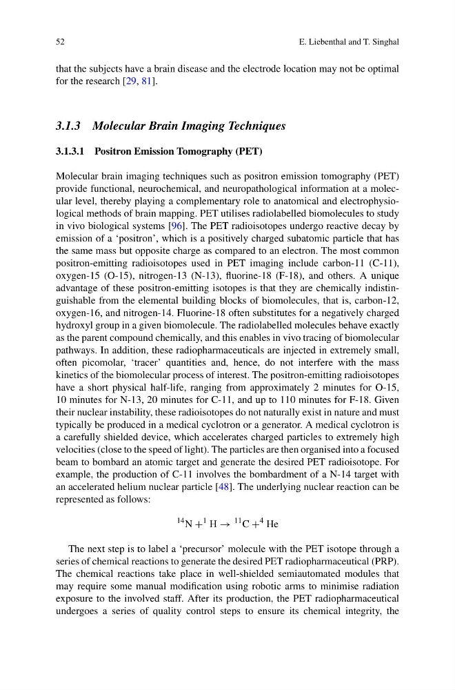
an accelerated helium nuclear particle [48]. The underlying nuclear reaction can be
represented as follows:
14 N +
1 H →
11 C +
4 He
The next step is to label a ‘precursor’ molecule with the PET isotope through a
series of chemical reactions to generate the desired PET radiopharmaceutical (PRP).
The chemical reactions take place in well-shielded semiautomated modules that
may require some manual modification using robotic arms to minimise radiation
exposure to the involved staff. After its production, the PET radiopharmaceutical
undergoes a series of quality control steps to ensure its chemical integrity, the
E. Liebenthal and T. Singhal
that the subjects have a brain disease and the electrode location may not be optimal
for the research [29, 81].
3.1.3 Molecular Brain Imaging Techniques
3.1.3.1 Positron Emission Tomography (PET)
Molecular brain imaging techniques such as positron emission tomography (PET)
provide functional, neurochemical, and neuropathological information at a molecular level, thereby playing a complementary role to anatomical and electrophysiological methods of brain mapping. PET utilises radiolabelled biomolecules to study
in vivo biological systems [96]. The PET radioisotopes undergo reactive decay by
emission of a ‘positron’, which is a positively charged subatomic particle that has
the same mass but opposite charge as compared to an electron. The most common
positron-emitting radioisotopes used in PET imaging include carbon-11 (C-11),
oxygen-15 (O-15), nitrogen-13 (N-13), fluorine-18 (F-18), and others. A unique
advantage of these positron-emitting isotopes is that they are chemically indistinguishable from the elemental building blocks of biomolecules, that is, carbon-12,
oxygen-16, and nitrogen-14. Fluorine-18 often substitutes for a negatively charged
hydroxyl group in a given biomolecule. The radiolabelled molecules behave exactly
as the parent compound chemically, and this enables in vivo tracing of biomolecular
pathways. In addition, these radiopharmaceuticals are injected in extremely small,
often picomolar, ‘tracer’ quantities and, hence, do not interfere with the mass
kinetics of the biomolecular process of interest. The positron-emitting radioisotopes
have a short physical half-life, ranging from approximately 2 minutes for O-15,
10 minutes for N-13, 20 minutes for C-11, and up to 110 minutes for F-18. Given
their nuclear instability, these radioisotopes do not naturally exist in nature and must
typically be produced in a medical cyclotron or a generator. A medical cyclotron is
a carefully shielded device, which accelerates charged particles to extremely high
velocities (close to the speed of light). The particles are then organised into a focused
beam to bombard an atomic target and generate the desired PET radioisotope. For
example, the production of C-11 involves the bombardment of a N-14 target with
an accelerated helium nuclear particle [48]. The underlying nuclear reaction can be
represented as follows:
14 N +
1 H →
11 C +
4 He
The next step is to label a ‘precursor’ molecule with the PET isotope through a
series of chemical reactions to generate the desired PET radiopharmaceutical (PRP).
The chemical reactions take place in well-shielded semiautomated modules that
may require some manual modification using robotic arms to minimise radiation
exposure to the involved staff. After its production, the PET radiopharmaceutical
undergoes a series of quality control steps to ensure its chemical integrity, the