

2 Human Brain Anatomy in 3D
35
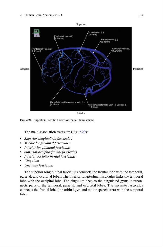
Fig. 2.24 Superficial cerebral veins of the left hemisphere
The main association tracts are (Fig. 2.29):
• Superior longitudinal fasciculus
• Middle longitudinal fasciculus
• Inferior longitudinal fasciculus
• Superior occipito-frontal fasciculus
• Inferior occipito-frontal fasciculus
• Cingulum
• Uncinate fasciculus
The superior longitudinal fasciculus connects the frontal lobe with the temporal,
parietal, and occipital lobes. The inferior longitudinal fasciculus links the temporal
lobe with the occipital lobe. The cingulum deep to the cingulated gyrus interconnects parts of the temporal, parietal, and occipital lobes. The uncinate fasciculus
connects the frontal lobe (the orbital gyri and motor speech area) with the temporal
lobe.
35
Fig. 2.24 Superficial cerebral veins of the left hemisphere
The main association tracts are (Fig. 2.29):
• Superior longitudinal fasciculus
• Middle longitudinal fasciculus
• Inferior longitudinal fasciculus
• Superior occipito-frontal fasciculus
• Inferior occipito-frontal fasciculus
• Cingulum
• Uncinate fasciculus
The superior longitudinal fasciculus connects the frontal lobe with the temporal,
parietal, and occipital lobes. The inferior longitudinal fasciculus links the temporal
lobe with the occipital lobe. The cingulum deep to the cingulated gyrus interconnects parts of the temporal, parietal, and occipital lobes. The uncinate fasciculus
connects the frontal lobe (the orbital gyri and motor speech area) with the temporal
lobe.