

328
H. Courtecuisse et al.
13.3 Example of Clinical Application: Constraint-Based
Simulation During Tumour Resection
This section introduces our approach to compensate for the brain shift induced
by the dura mater opening [8]. The method combines a biomechanical model
build from pre-operative images and image-based constraints extracted from intraoperative US.
Before surgery, a patient-specific finite element (FE) model of the brain is
constructed from pre-operative MRI images. This model accounts for the soft
tissue morphology but also vessels located around the tumour. During surgery, after
opening, the skull and dura mater, localised Doppler, and B-mode ultrasound images
were acquired directly in contact with the brain. The vascular tree and the footprint
of the ultrasound probe are then extracted from these intra-operative images.
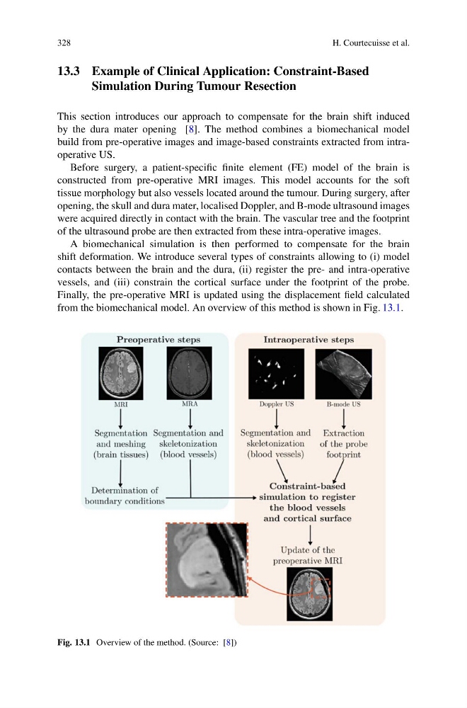
A biomechanical simulation is then performed to compensate for the brain
shift deformation. We introduce several types of constraints allowing to (i) model
contacts between the brain and the dura, (ii) register the pre- and intra-operative
vessels, and (iii) constrain the cortical surface under the footprint of the probe.
Finally, the pre-operative MRI is updated using the displacement field calculated
from the biomechanical model. An overview of this method is shown in Fig. 13.1.
Fig. 13.1 Overview of the method. (Source: [8])
H. Courtecuisse et al.
13.3 Example of Clinical Application: Constraint-Based
Simulation During Tumour Resection
This section introduces our approach to compensate for the brain shift induced
by the dura mater opening [8]. The method combines a biomechanical model
build from pre-operative images and image-based constraints extracted from intraoperative US.
Before surgery, a patient-specific finite element (FE) model of the brain is
constructed from pre-operative MRI images. This model accounts for the soft
tissue morphology but also vessels located around the tumour. During surgery, after
opening, the skull and dura mater, localised Doppler, and B-mode ultrasound images
were acquired directly in contact with the brain. The vascular tree and the footprint
of the ultrasound probe are then extracted from these intra-operative images.
A biomechanical simulation is then performed to compensate for the brain
shift deformation. We introduce several types of constraints allowing to (i) model
contacts between the brain and the dura, (ii) register the pre- and intra-operative
vessels, and (iii) constrain the cortical surface under the footprint of the probe.
Finally, the pre-operative MRI is updated using the displacement field calculated
from the biomechanical model. An overview of this method is shown in Fig. 13.1.
Fig. 13.1 Overview of the method. (Source: [8])