

13 Computational Biomechanics of the Brain in the Operating Theatre
323
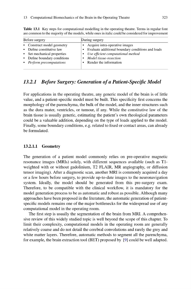
Table 13.1 Key steps for computational modelling in the operating theatre. Terms in regular font
are common to the majority of the models, while ones in italic could be considered for improvement
Before surgery
During surgery
• Construct model geometry
• Define constitutive law
• Set mechanical properties
• Define boundary conditions
• Perform precomputations
• Acquire intra-operative images
• Evaluate additional boundary conditions and loads
• Use efficient computational method
• Model tissue-resection
• Render the information
13.2.1 Before Surgery: Generation of a Patient-Specific Model
For applications in the operating theatre, any generic model of the brain is of little
value, and a patient-specific model must be built. This specificity first concerns the
morphology of the parenchyma, the bulk of the model, and the inner structures such
as the dura mater, ventricles, or tumour, if any. While the constitutive law of the
brain tissue is usually generic, estimating the patient’s own rheological parameters
could be a valuable addition, depending on the type of loads applied to the model.
Finally, some boundary conditions, e.g. related to fixed or contact areas, can already
be formulated.
13.2.1.1 Geometry
The generation of a patient model commonly relies on pre-operative magnetic
resonance images (MRIs) solely, with different sequences available (such as T1weighted with or without gadolinium, T2 FLAIR, MR angiography, or diffusion
tensor imaging). After a diagnostic scan, another MRI is commonly acquired a day
or a few hours before surgery, to provide up-to-date images to the neuronavigation
system. Ideally, the model should be generated from this pre-surgery exam.
Therefore, to be compatible with the clinical workflow, it is mandatory for the
model generation process to be as automatic and robust as possible. Although many
approaches have been proposed in the literature, the automatic generation of patientspecific models remains one of the major bottlenecks for the widespread use of any
computational model in the operating room.
The first step is usually the segmentation of the brain from MRI. A comprehensive review of this widely studied topic is well beyond the scope of this chapter. To
limit their complexity, computational models in the operating room are generally
relatively coarse and do not detail the cerebral convolutions and rarely the grey and
white matter layers. Therefore, automatic methods to segment all the parenchyma,
for example, the brain extraction tool (BET) proposed by [9] could be well adapted.
323
Table 13.1 Key steps for computational modelling in the operating theatre. Terms in regular font
are common to the majority of the models, while ones in italic could be considered for improvement
Before surgery
During surgery
• Construct model geometry
• Define constitutive law
• Set mechanical properties
• Define boundary conditions
• Perform precomputations
• Acquire intra-operative images
• Evaluate additional boundary conditions and loads
• Use efficient computational method
• Model tissue-resection
• Render the information
13.2.1 Before Surgery: Generation of a Patient-Specific Model
For applications in the operating theatre, any generic model of the brain is of little
value, and a patient-specific model must be built. This specificity first concerns the
morphology of the parenchyma, the bulk of the model, and the inner structures such
as the dura mater, ventricles, or tumour, if any. While the constitutive law of the
brain tissue is usually generic, estimating the patient’s own rheological parameters
could be a valuable addition, depending on the type of loads applied to the model.
Finally, some boundary conditions, e.g. related to fixed or contact areas, can already
be formulated.
13.2.1.1 Geometry
The generation of a patient model commonly relies on pre-operative magnetic
resonance images (MRIs) solely, with different sequences available (such as T1weighted with or without gadolinium, T2 FLAIR, MR angiography, or diffusion
tensor imaging). After a diagnostic scan, another MRI is commonly acquired a day
or a few hours before surgery, to provide up-to-date images to the neuronavigation
system. Ideally, the model should be generated from this pre-surgery exam.
Therefore, to be compatible with the clinical workflow, it is mandatory for the
model generation process to be as automatic and robust as possible. Although many
approaches have been proposed in the literature, the automatic generation of patientspecific models remains one of the major bottlenecks for the widespread use of any
computational model in the operating room.
The first step is usually the segmentation of the brain from MRI. A comprehensive review of this widely studied topic is well beyond the scope of this chapter. To
limit their complexity, computational models in the operating room are generally
relatively coarse and do not detail the cerebral convolutions and rarely the grey and
white matter layers. Therefore, automatic methods to segment all the parenchyma,
for example, the brain extraction tool (BET) proposed by [9] could be well adapted.