

9 Modelling of Cerebrospinal Fluid Flow by Computational Fluid Dynamics
227
9.3.2 Subarachnoid Space
A number of CFD models have been applied to elucidate CSF dynamics in the SAS.
These models can be roughly divided into studies investigating (a) CSF flow under
normal physiologic conditions, (b) syringomyelia, (c) Chiari malformation and (d)
intrathecal drug delivery and solute transport. A review of spinal SAS CFD studies
was provided by Khani et al. in 2018 [69], a review of syringomyelia biomechanics
was provided by Elliott et al. in 2013 [70], and a review of Chiari malformation
biomechanics was provided by Shaffer et al. in 2011 [71]. An example of spinal
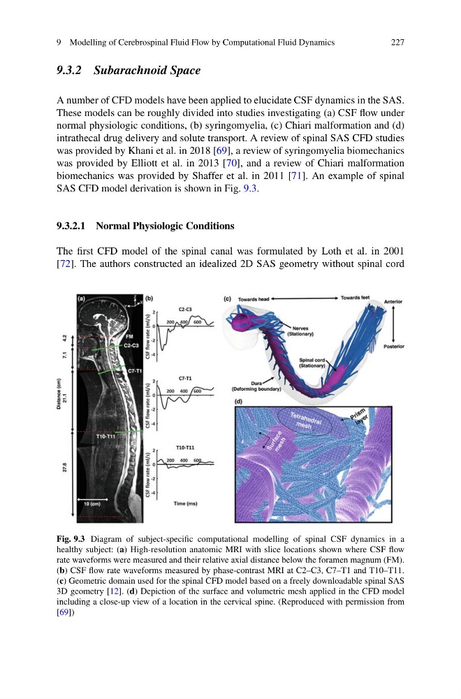
SAS CFD model derivation is shown in Fig. 9.3.
9.3.2.1 Normal Physiologic Conditions
The first CFD model of the spinal canal was formulated by Loth et al. in 2001
[72]. The authors constructed an idealized 2D SAS geometry without spinal cord
Fig. 9.3 Diagram of subject-specific computational modelling of spinal CSF dynamics in a
healthy subject: (a) High-resolution anatomic MRI with slice locations shown where CSF flow
rate waveforms were measured and their relative axial distance below the foramen magnum (FM).
(b) CSF flow rate waveforms measured by phase-contrast MRI at C2–C3, C7–T1 and T10–T11.
(c) Geometric domain used for the spinal CFD model based on a freely downloadable spinal SAS
3D geometry [12]. (d) Depiction of the surface and volumetric mesh applied in the CFD model
including a close-up view of a location in the cervical spine. (Reproduced with permission from
[69])
227
9.3.2 Subarachnoid Space
A number of CFD models have been applied to elucidate CSF dynamics in the SAS.
These models can be roughly divided into studies investigating (a) CSF flow under
normal physiologic conditions, (b) syringomyelia, (c) Chiari malformation and (d)
intrathecal drug delivery and solute transport. A review of spinal SAS CFD studies
was provided by Khani et al. in 2018 [69], a review of syringomyelia biomechanics
was provided by Elliott et al. in 2013 [70], and a review of Chiari malformation
biomechanics was provided by Shaffer et al. in 2011 [71]. An example of spinal
SAS CFD model derivation is shown in Fig. 9.3.
9.3.2.1 Normal Physiologic Conditions
The first CFD model of the spinal canal was formulated by Loth et al. in 2001
[72]. The authors constructed an idealized 2D SAS geometry without spinal cord
Fig. 9.3 Diagram of subject-specific computational modelling of spinal CSF dynamics in a
healthy subject: (a) High-resolution anatomic MRI with slice locations shown where CSF flow
rate waveforms were measured and their relative axial distance below the foramen magnum (FM).
(b) CSF flow rate waveforms measured by phase-contrast MRI at C2–C3, C7–T1 and T10–T11.
(c) Geometric domain used for the spinal CFD model based on a freely downloadable spinal SAS
3D geometry [12]. (d) Depiction of the surface and volumetric mesh applied in the CFD model
including a close-up view of a location in the cervical spine. (Reproduced with permission from
[69])