

2 Human Brain Anatomy in 3D
11
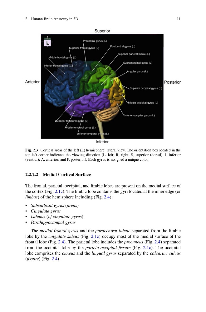
Fig. 2.3 Cortical areas of the left (L) hemisphere: lateral view. The orientation box located in the
top-left corner indicates the viewing direction (L, left; R, right; S, superior (dorsal); I, inferior
(ventral); A, anterior; and P, posterior). Each gyrus is assigned a unique color
2.2.2.2 Medial Cortical Surface
The frontal, parietal, occipital, and limbic lobes are present on the medial surface of
the cortex (Fig. 2.1c). The limbic lobe contains the gyri located at the inner edge (or
limbus) of the hemisphere including (Fig. 2.4):
• Subcallosal gyrus (areas)
• Cingulate gyrus
• Isthmus (of cingulate gyrus)
• Parahippocampal gyrus
The medial frontal gyrus and the paracentral lobule separated from the limbic
lobe by the cingulate sulcus (Fig. 2.1c) occupy most of the medial surface of the
frontal lobe (Fig. 2.4). The parietal lobe includes the precuneus (Fig. 2.4) separated
from the occipital lobe by the parieto-occipital fissure (Fig. 2.1c). The occipital
lobe comprises the cuneus and the lingual gyrus separated by the calcarine sulcus
(fissure) (Fig. 2.4).
11
Fig. 2.3 Cortical areas of the left (L) hemisphere: lateral view. The orientation box located in the
top-left corner indicates the viewing direction (L, left; R, right; S, superior (dorsal); I, inferior
(ventral); A, anterior; and P, posterior). Each gyrus is assigned a unique color
2.2.2.2 Medial Cortical Surface
The frontal, parietal, occipital, and limbic lobes are present on the medial surface of
the cortex (Fig. 2.1c). The limbic lobe contains the gyri located at the inner edge (or
limbus) of the hemisphere including (Fig. 2.4):
• Subcallosal gyrus (areas)
• Cingulate gyrus
• Isthmus (of cingulate gyrus)
• Parahippocampal gyrus
The medial frontal gyrus and the paracentral lobule separated from the limbic
lobe by the cingulate sulcus (Fig. 2.1c) occupy most of the medial surface of the
frontal lobe (Fig. 2.4). The parietal lobe includes the precuneus (Fig. 2.4) separated
from the occipital lobe by the parieto-occipital fissure (Fig. 2.1c). The occipital
lobe comprises the cuneus and the lingual gyrus separated by the calcarine sulcus
(fissure) (Fig. 2.4).