

7 Biomechanical Modelling of the Brain for Neuronavigation in Epilepsy Surgery
167
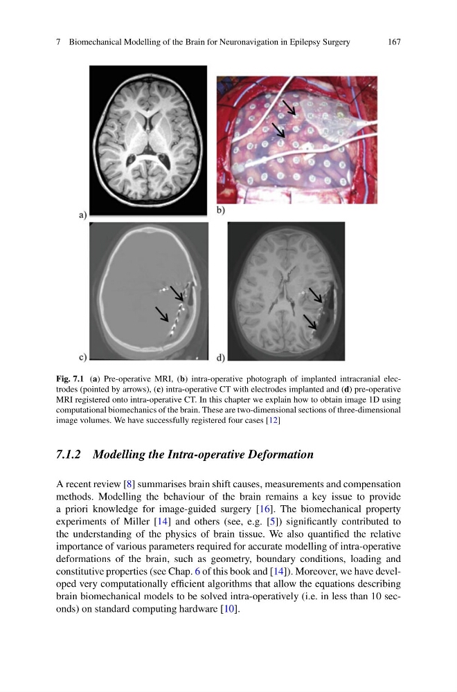
Fig. 7.1 (a) Pre-operative MRI, (b) intra-operative photograph of implanted intracranial electrodes (pointed by arrows), (c) intra-operative CT with electrodes implanted and (d) pre-operative
MRI registered onto intra-operative CT. In this chapter we explain how to obtain image 1D using
computational biomechanics of the brain. These are two-dimensional sections of three-dimensional
image volumes. We have successfully registered four cases [12]
7.1.2 Modelling the Intra-operative Deformation
A recent review [8] summarises brain shift causes, measurements and compensation
methods. Modelling the behaviour of the brain remains a key issue to provide
a priori knowledge for image-guided surgery [16]. The biomechanical property
experiments of Miller [14] and others (see, e.g. [5]) significantly contributed to
the understanding of the physics of brain tissue. We also quantified the relative
importance of various parameters required for accurate modelling of intra-operative
deformations of the brain, such as geometry, boundary conditions, loading and
constitutive properties (see Chap. 6 of this book and [14]). Moreover, we have developed very computationally efficient algorithms that allow the equations describing
brain biomechanical models to be solved intra-operatively (i.e. in less than 10 seconds) on standard computing hardware [10].
167
Fig. 7.1 (a) Pre-operative MRI, (b) intra-operative photograph of implanted intracranial electrodes (pointed by arrows), (c) intra-operative CT with electrodes implanted and (d) pre-operative
MRI registered onto intra-operative CT. In this chapter we explain how to obtain image 1D using
computational biomechanics of the brain. These are two-dimensional sections of three-dimensional
image volumes. We have successfully registered four cases [12]
7.1.2 Modelling the Intra-operative Deformation
A recent review [8] summarises brain shift causes, measurements and compensation
methods. Modelling the behaviour of the brain remains a key issue to provide
a priori knowledge for image-guided surgery [16]. The biomechanical property
experiments of Miller [14] and others (see, e.g. [5]) significantly contributed to
the understanding of the physics of brain tissue. We also quantified the relative
importance of various parameters required for accurate modelling of intra-operative
deformations of the brain, such as geometry, boundary conditions, loading and
constitutive properties (see Chap. 6 of this book and [14]). Moreover, we have developed very computationally efficient algorithms that allow the equations describing
brain biomechanical models to be solved intra-operatively (i.e. in less than 10 seconds) on standard computing hardware [10].