

150
K. Miller et al.
6.3.1 Generation of Computational Grids: From Medical
Images to Finite Element Meshes
Pre-operative and intra-operative medical image datasets of 33 patients with cerebral
gliomas were randomly selected from a retrospective database of 859 intracranial
tumour cases available at Boston Children’s Hospital [94]. Imaging was performed
using a 0.5 T open MR system in the neurosurgical suite. The resolution of
the images is 0.85 × 0.85 × 2.5 mm 3 . Consent was obtained for the use of
the anonymised retrospective image database, in accordance with the Institutional
Review Board of Boston Children’s Hospital.
A three-dimensional (3D) surface model of each patient’s brain was created
from segmented pre-operative magnetic resonance images (MRIs). Following our
previous studies on predicting craniotomy-induced deformations within the brain
[11, 95–98], in this investigation, different material properties were assigned to
the parenchyma, tumour and ventricles. Accordingly, to obtain the information for
building the computational grids (finite element meshes), the parenchyma, tumour
and ventricles were segmented using the region growing algorithm implemented in
3D Slicer, followed by manual correction.
The meshes were constructed using low-order elements (linear tetrahedron
or hexahedron) to meet the requirement that computations be conducted intraoperatively, i.e. within no more than ca. 1 min. To prevent volumetric locking,
tetrahedral elements with average nodal pressure (ANP) formulation were used [99].
The meshes were generated using IA-FEMesh [100] and HyperMesh (commercial
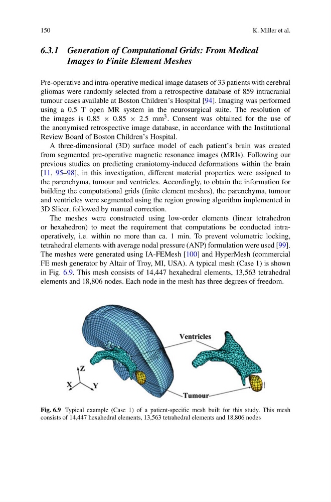
FE mesh generator by Altair of Troy, MI, USA). A typical mesh (Case 1) is shown
in Fig. 6.9. This mesh consists of 14,447 hexahedral elements, 13,563 tetrahedral
elements and 18,806 nodes. Each node in the mesh has three degrees of freedom.
Fig. 6.9 Typical example (Case 1) of a patient-specific mesh built for this study. This mesh
consists of 14,447 hexahedral elements, 13,563 tetrahedral elements and 18,806 nodes
K. Miller et al.
6.3.1 Generation of Computational Grids: From Medical
Images to Finite Element Meshes
Pre-operative and intra-operative medical image datasets of 33 patients with cerebral
gliomas were randomly selected from a retrospective database of 859 intracranial
tumour cases available at Boston Children’s Hospital [94]. Imaging was performed
using a 0.5 T open MR system in the neurosurgical suite. The resolution of
the images is 0.85 × 0.85 × 2.5 mm 3 . Consent was obtained for the use of
the anonymised retrospective image database, in accordance with the Institutional
Review Board of Boston Children’s Hospital.
A three-dimensional (3D) surface model of each patient’s brain was created
from segmented pre-operative magnetic resonance images (MRIs). Following our
previous studies on predicting craniotomy-induced deformations within the brain
[11, 95–98], in this investigation, different material properties were assigned to
the parenchyma, tumour and ventricles. Accordingly, to obtain the information for
building the computational grids (finite element meshes), the parenchyma, tumour
and ventricles were segmented using the region growing algorithm implemented in
3D Slicer, followed by manual correction.
The meshes were constructed using low-order elements (linear tetrahedron
or hexahedron) to meet the requirement that computations be conducted intraoperatively, i.e. within no more than ca. 1 min. To prevent volumetric locking,
tetrahedral elements with average nodal pressure (ANP) formulation were used [99].
The meshes were generated using IA-FEMesh [100] and HyperMesh (commercial
FE mesh generator by Altair of Troy, MI, USA). A typical mesh (Case 1) is shown
in Fig. 6.9. This mesh consists of 14,447 hexahedral elements, 13,563 tetrahedral
elements and 18,806 nodes. Each node in the mesh has three degrees of freedom.
Fig. 6.9 Typical example (Case 1) of a patient-specific mesh built for this study. This mesh
consists of 14,447 hexahedral elements, 13,563 tetrahedral elements and 18,806 nodes