

4.1 Introduction
69
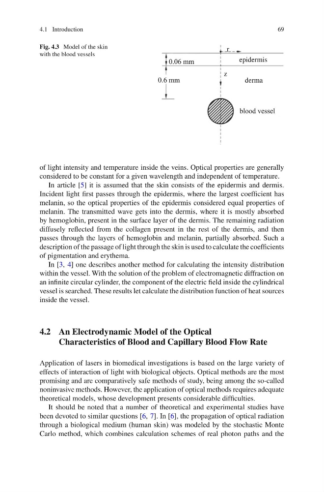
Fig. 4.3 Model of the skin
with the blood vessels
derma
epidermis
0.6 mm
z
r
of light intensity and temperature inside the veins. Optical properties are generally
considered to be constant for a given wavelength and independent of temperature.
In article [5] it is assumed that the skin consists of the epidermis and dermis.
Incident light first passes through the epidermis, where the largest coefficient has
melanin, so the optical properties of the epidermis considered equal properties of
melanin. The transmitted wave gets into the dermis, where it is mostly absorbed
by hemoglobin, present in the surface layer of the dermis. The remaining radiation
diffusely reflected from the collagen present in the rest of the dermis, and then
passes through the layers of hemoglobin and melanin, partially absorbed. Such a
description of the passage of light through the skin is used to calculate the coefficients
of pigmentation and erythema.
In [3, 4] one describes another method for calculating the intensity distribution
within the vessel. With the solution of the problem of electromagnetic diffraction on
an infinite circular cylinder, the component of the electric field inside the cylindrical
vessel is searched. These results let calculate the distribution function of heat sources
inside the vessel.
4.2 An Electrodynamic Model of the Optical
Characteristics of Blood and Capillary Blood Flow Rate
Application of lasers in biomedical investigations is based on the large variety of
effects of interaction of light with biological objects. Optical methods are the most
promising and are comparatively safe methods of study, being among the so-called
noninvasive methods. However, the application of optical methods requires adequate
theoretical models, whose development presents considerable difficulties.
It should be noted that a number of theoretical and experimental studies have
been devoted to similar questions [6, 7]. In [6], the propagation of optical radiation
through a biological medium (human skin) was modeled by the stochastic Monte
Carlo method, which combines calculation schemes of real photon paths and the
69
Fig. 4.3 Model of the skin
with the blood vessels
derma
epidermis
0.6 mm
z
r
of light intensity and temperature inside the veins. Optical properties are generally
considered to be constant for a given wavelength and independent of temperature.
In article [5] it is assumed that the skin consists of the epidermis and dermis.
Incident light first passes through the epidermis, where the largest coefficient has
melanin, so the optical properties of the epidermis considered equal properties of
melanin. The transmitted wave gets into the dermis, where it is mostly absorbed
by hemoglobin, present in the surface layer of the dermis. The remaining radiation
diffusely reflected from the collagen present in the rest of the dermis, and then
passes through the layers of hemoglobin and melanin, partially absorbed. Such a
description of the passage of light through the skin is used to calculate the coefficients
of pigmentation and erythema.
In [3, 4] one describes another method for calculating the intensity distribution
within the vessel. With the solution of the problem of electromagnetic diffraction on
an infinite circular cylinder, the component of the electric field inside the cylindrical
vessel is searched. These results let calculate the distribution function of heat sources
inside the vessel.
4.2 An Electrodynamic Model of the Optical
Characteristics of Blood and Capillary Blood Flow Rate
Application of lasers in biomedical investigations is based on the large variety of
effects of interaction of light with biological objects. Optical methods are the most
promising and are comparatively safe methods of study, being among the so-called
noninvasive methods. However, the application of optical methods requires adequate
theoretical models, whose development presents considerable difficulties.
It should be noted that a number of theoretical and experimental studies have
been devoted to similar questions [6, 7]. In [6], the propagation of optical radiation
through a biological medium (human skin) was modeled by the stochastic Monte
Carlo method, which combines calculation schemes of real photon paths and the