

62
3 Study of Electrophysical Characteristics of Blood …
Then,
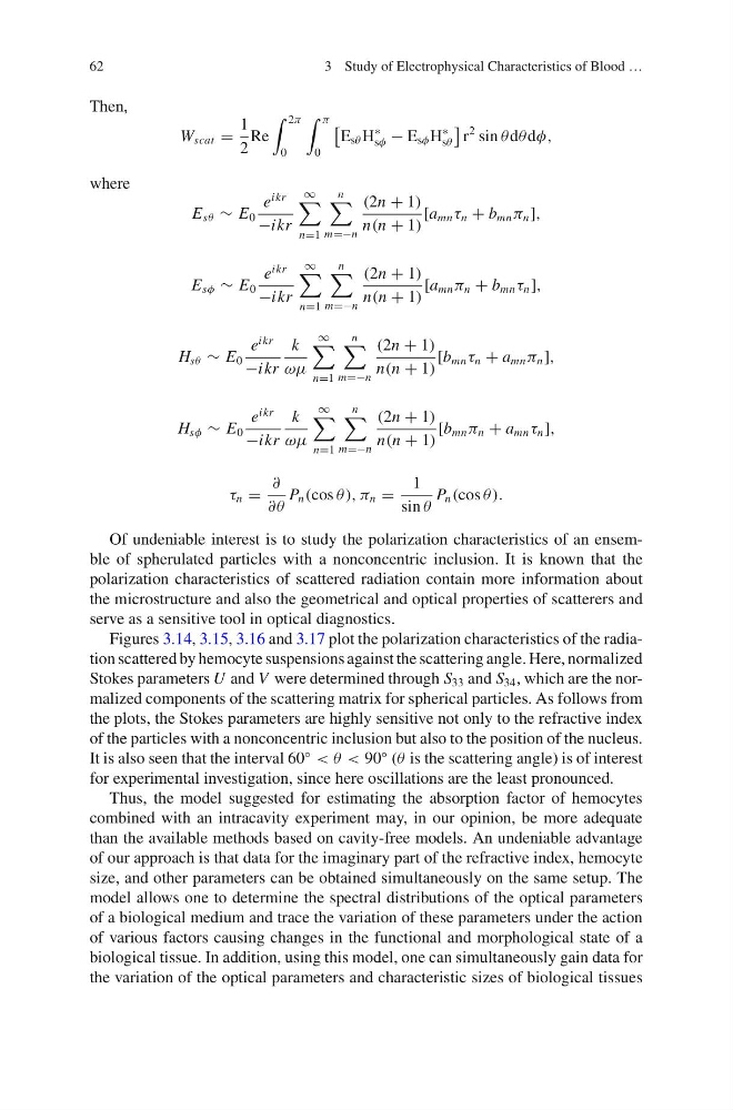
W scat =
1
2
Re
2π
0
π
0
E sθ H
∗
sφ − E sφ H
∗
sθ
r
2 sin θ dθ dφ,
where
E sθ ∼ E 0
e
ikr
−ikr
∞
n=1
n
m=−n
(2n + 1)
n(n + 1)
[a mn τ n + b mn π n ],
E sφ ∼ E 0
e
ikr
−ikr
∞
n=1
n
m=−n
(2n + 1)
n(n + 1)
[a mn π n + b mn τ n ],
H sθ ∼ E 0
e
ikr
−ikr
k
ωμ
∞
n=1
n
m=−n
(2n + 1)
n(n + 1)
[b mn τ n + a mn π n ],
H sφ ∼ E 0
e
ikr
−ikr
k
ωμ
∞
n=1
n
m=−n
(2n + 1)
n(n + 1)
[b mn π n + a mn τ n ],
τ n =
∂
∂θ
P n (cos θ), π n =
1
sin θ
P n (cos θ).
Of undeniable interest is to study the polarization characteristics of an ensemble of spherulated particles with a nonconcentric inclusion. It is known that the
polarization characteristics of scattered radiation contain more information about
the microstructure and also the geometrical and optical properties of scatterers and
serve as a sensitive tool in optical diagnostics.
Figures 3.14, 3.15, 3.16 and 3.17 plot the polarization characteristics of the radiation scattered by hemocyte suspensions against the scattering angle. Here, normalized
Stokes parameters U and V were determined through S 33 and S 34 , which are the normalized components of the scattering matrix for spherical particles. As follows from
the plots, the Stokes parameters are highly sensitive not only to the refractive index
of the particles with a nonconcentric inclusion but also to the position of the nucleus.
It is also seen that the interval 60
◦
< θ < 90
◦ (θ is the scattering angle) is of interest
for experimental investigation, since here oscillations are the least pronounced.
Thus, the model suggested for estimating the absorption factor of hemocytes
combined with an intracavity experiment may, in our opinion, be more adequate
than the available methods based on cavity-free models. An undeniable advantage
of our approach is that data for the imaginary part of the refractive index, hemocyte
size, and other parameters can be obtained simultaneously on the same setup. The
model allows one to determine the spectral distributions of the optical parameters
of a biological medium and trace the variation of these parameters under the action
of various factors causing changes in the functional and morphological state of a
biological tissue. In addition, using this model, one can simultaneously gain data for
the variation of the optical parameters and characteristic sizes of biological tissues
3 Study of Electrophysical Characteristics of Blood …
Then,
W scat =
1
2
Re
2π
0
π
0
E sθ H
∗
sφ − E sφ H
∗
sθ
r
2 sin θ dθ dφ,
where
E sθ ∼ E 0
e
ikr
−ikr
∞
n=1
n
m=−n
(2n + 1)
n(n + 1)
[a mn τ n + b mn π n ],
E sφ ∼ E 0
e
ikr
−ikr
∞
n=1
n
m=−n
(2n + 1)
n(n + 1)
[a mn π n + b mn τ n ],
H sθ ∼ E 0
e
ikr
−ikr
k
ωμ
∞
n=1
n
m=−n
(2n + 1)
n(n + 1)
[b mn τ n + a mn π n ],
H sφ ∼ E 0
e
ikr
−ikr
k
ωμ
∞
n=1
n
m=−n
(2n + 1)
n(n + 1)
[b mn π n + a mn τ n ],
τ n =
∂
∂θ
P n (cos θ), π n =
1
sin θ
P n (cos θ).
Of undeniable interest is to study the polarization characteristics of an ensemble of spherulated particles with a nonconcentric inclusion. It is known that the
polarization characteristics of scattered radiation contain more information about
the microstructure and also the geometrical and optical properties of scatterers and
serve as a sensitive tool in optical diagnostics.
Figures 3.14, 3.15, 3.16 and 3.17 plot the polarization characteristics of the radiation scattered by hemocyte suspensions against the scattering angle. Here, normalized
Stokes parameters U and V were determined through S 33 and S 34 , which are the normalized components of the scattering matrix for spherical particles. As follows from
the plots, the Stokes parameters are highly sensitive not only to the refractive index
of the particles with a nonconcentric inclusion but also to the position of the nucleus.
It is also seen that the interval 60
◦
< θ < 90
◦ (θ is the scattering angle) is of interest
for experimental investigation, since here oscillations are the least pronounced.
Thus, the model suggested for estimating the absorption factor of hemocytes
combined with an intracavity experiment may, in our opinion, be more adequate
than the available methods based on cavity-free models. An undeniable advantage
of our approach is that data for the imaginary part of the refractive index, hemocyte
size, and other parameters can be obtained simultaneously on the same setup. The
model allows one to determine the spectral distributions of the optical parameters
of a biological medium and trace the variation of these parameters under the action
of various factors causing changes in the functional and morphological state of a
biological tissue. In addition, using this model, one can simultaneously gain data for
the variation of the optical parameters and characteristic sizes of biological tissues