

1.3 Structure and Optical Properties of Blood
7
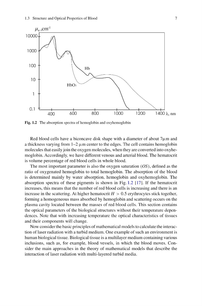
Fig. 1.2 The absorption spectra of hemoglobin and oxyhemoglobin
Red blood cells have a biconcave disk shape with a diameter of about 7µm and
a thickness varying from 1–2 µm center to the edges. The cell contains hemoglobin
molecules that easily join the oxygen molecules, when they are converted into oxyhemoglobin. Accordingly, we have different venous and arterial blood. The hematocrit
is volume percentage of red blood cells in whole blood.
The most important parameter is also the oxygen saturation (OS), defined as the
ratio of oxygenated hemoglobin to total hemoglobin. The absorption of the blood
is determined mainly by water absorption, hemoglobin and oxyhemoglobin. The
absorption spectra of these pigments is shown in Fig. 1.2 [17]. If the hematocrit
increases, this means that the number of red blood cells is increasing and there is an
increase in the scattering. At higher hematocrit H > 0.5 erythrocytes stick together,
forming a homogeneous mass absorbed by hemoglobin and scattering occurs on the
plasma cavity located between the masses of red blood cells. This section contains
the optical parameters of the biological structures without their temperature dependences. Note that with increasing temperature the optical characteristics of tissues
and their components will change.
Now consider the basic principles of mathematical models to calculate the interaction of laser radiation with a turbid medium. One example of such an environment is
human biological tissue. Biological tissue is a multilayer medium containing various
inclusions, such as, for example, blood vessels, in which the blood moves. Consider the main approaches in the theory of mathematical models that describe the
interaction of laser radiation with multi-layered turbid media.
7
Fig. 1.2 The absorption spectra of hemoglobin and oxyhemoglobin
Red blood cells have a biconcave disk shape with a diameter of about 7µm and
a thickness varying from 1–2 µm center to the edges. The cell contains hemoglobin
molecules that easily join the oxygen molecules, when they are converted into oxyhemoglobin. Accordingly, we have different venous and arterial blood. The hematocrit
is volume percentage of red blood cells in whole blood.
The most important parameter is also the oxygen saturation (OS), defined as the
ratio of oxygenated hemoglobin to total hemoglobin. The absorption of the blood
is determined mainly by water absorption, hemoglobin and oxyhemoglobin. The
absorption spectra of these pigments is shown in Fig. 1.2 [17]. If the hematocrit
increases, this means that the number of red blood cells is increasing and there is an
increase in the scattering. At higher hematocrit H > 0.5 erythrocytes stick together,
forming a homogeneous mass absorbed by hemoglobin and scattering occurs on the
plasma cavity located between the masses of red blood cells. This section contains
the optical parameters of the biological structures without their temperature dependences. Note that with increasing temperature the optical characteristics of tissues
and their components will change.
Now consider the basic principles of mathematical models to calculate the interaction of laser radiation with a turbid medium. One example of such an environment is
human biological tissue. Biological tissue is a multilayer medium containing various
inclusions, such as, for example, blood vessels, in which the blood moves. Consider the main approaches in the theory of mathematical models that describe the
interaction of laser radiation with multi-layered turbid media.