

8.4 Numerical Calculations for a Model Medium and Conclusions
151
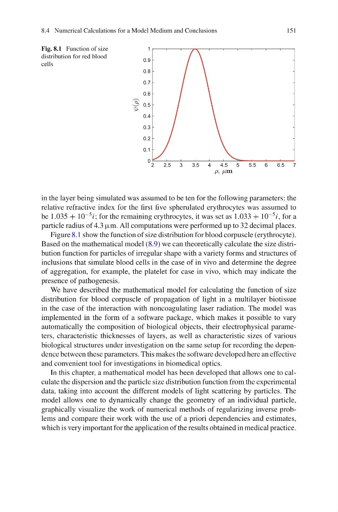
Fig. 8.1 Function of size
distribution for red blood
cells
2
2.5
3
3.5
4
4.5
5
5.5
6
6.5
7
0
0.1
0.2
0.3
0.4
0.5
0.6
0.7
0.8
0.9
1
in the layer being simulated was assumed to be ten for the following parameters: the
relative refractive index for the first five spherulated erythrocytes was assumed to
be 1.035 + 10
−5 i; for the remaining erythrocytes, it was set as 1.033 + 10
−5 i, for a
particle radius of 4.3 µm. All computations were performed up to 32 decimal places.
Figure 8.1 show the function of size distribution for blood corpuscle (erythrocyte).
Based on the mathematical model (8.9) we can theoretically calculate the size distribution function for particles of irregular shape with a variety forms and structures of
inclusions that simulate blood cells in the case of in vivo and determine the degree
of aggregation, for example, the platelet for case in vivo, which may indicate the
presence of pathogenesis.
We have described the mathematical model for calculating the function of size
distribution for blood corpuscle of propagation of light in a multilayer biotissue
in the case of the interaction with noncoagulating laser radiation. The model was
implemented in the form of a software package, which makes it possible to vary
automatically the composition of biological objects, their electrophysical parameters, characteristic thicknesses of layers, as well as characteristic sizes of various
biological structures under investigation on the same setup for recording the dependence between these parameters. This makes the software developed here an effective
and convenient tool for investigations in biomedical optics.
In this chapter, a mathematical model has been developed that allows one to calculate the dispersion and the particle size distribution function from the experimental
data, taking into account the different models of light scattering by particles. The
model allows one to dynamically change the geometry of an individual particle,
graphically visualize the work of numerical methods of regularizing inverse problems and compare their work with the use of a priori dependencies and estimates,
which is very important for the application of the results obtained in medical practice.
151
Fig. 8.1 Function of size
distribution for red blood
cells
2
2.5
3
3.5
4
4.5
5
5.5
6
6.5
7
0
0.1
0.2
0.3
0.4
0.5
0.6
0.7
0.8
0.9
1
in the layer being simulated was assumed to be ten for the following parameters: the
relative refractive index for the first five spherulated erythrocytes was assumed to
be 1.035 + 10
−5 i; for the remaining erythrocytes, it was set as 1.033 + 10
−5 i, for a
particle radius of 4.3 µm. All computations were performed up to 32 decimal places.
Figure 8.1 show the function of size distribution for blood corpuscle (erythrocyte).
Based on the mathematical model (8.9) we can theoretically calculate the size distribution function for particles of irregular shape with a variety forms and structures of
inclusions that simulate blood cells in the case of in vivo and determine the degree
of aggregation, for example, the platelet for case in vivo, which may indicate the
presence of pathogenesis.
We have described the mathematical model for calculating the function of size
distribution for blood corpuscle of propagation of light in a multilayer biotissue
in the case of the interaction with noncoagulating laser radiation. The model was
implemented in the form of a software package, which makes it possible to vary
automatically the composition of biological objects, their electrophysical parameters, characteristic thicknesses of layers, as well as characteristic sizes of various
biological structures under investigation on the same setup for recording the dependence between these parameters. This makes the software developed here an effective
and convenient tool for investigations in biomedical optics.
In this chapter, a mathematical model has been developed that allows one to calculate the dispersion and the particle size distribution function from the experimental
data, taking into account the different models of light scattering by particles. The
model allows one to dynamically change the geometry of an individual particle,
graphically visualize the work of numerical methods of regularizing inverse problems and compare their work with the use of a priori dependencies and estimates,
which is very important for the application of the results obtained in medical practice.