

7.4 Numerical Calculations for a Model Medium and Conclusions
141
In this case, the scattering cross section is defined a
C scat =
W scat
I i
,
where I i is incident intensity
W scat =
A
S scat · e r d A, S scat =
c
8π
Re[E
j
scat × H
j∗
scat ],
E
j
scat =
i
k
× × ×(e z U
j
scat ) + + × (e z V
j
scat ),
H
j
scat = −n o ×(e z U
j
scat ) +
i
k
× × ×(e z V
j
scat ),
U
j
scat , V
j
scat defined by the expression (7.18).
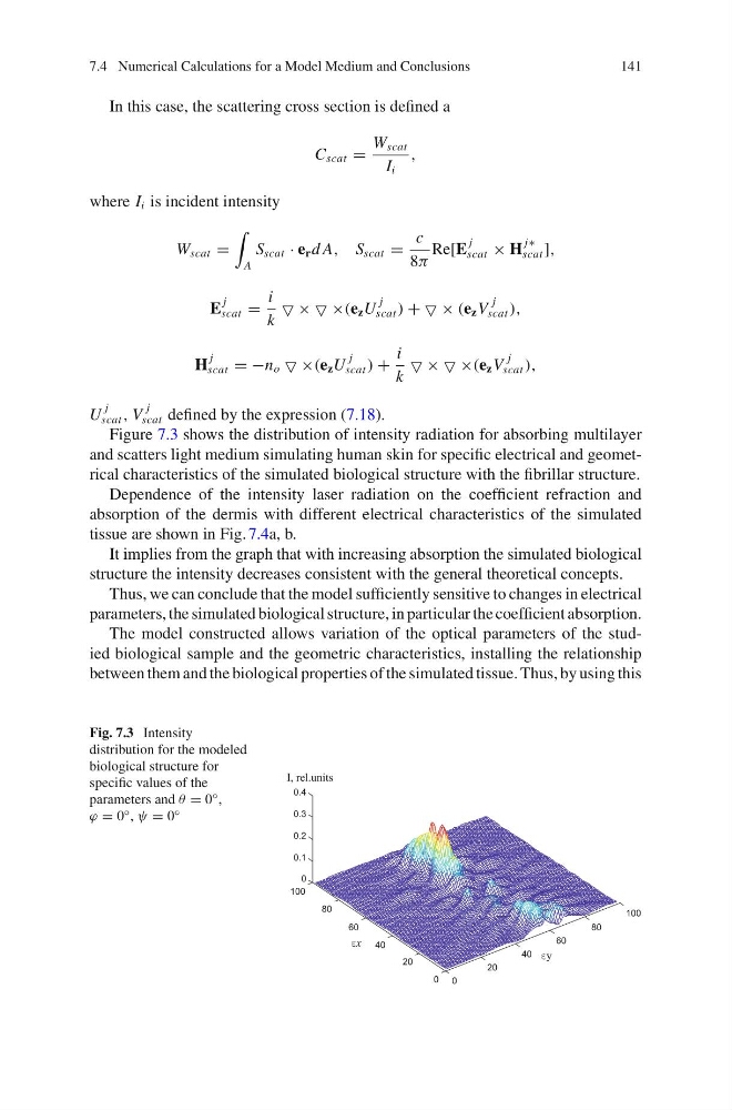
Figure 7.3 shows the distribution of intensity radiation for absorbing multilayer
and scatters light medium simulating human skin for specific electrical and geometrical characteristics of the simulated biological structure with the fibrillar structure.
Dependence of the intensity laser radiation on the coefficient refraction and
absorption of the dermis with different electrical characteristics of the simulated
tissue are shown in Fig. 7.4a, b.
It implies from the graph that with increasing absorption the simulated biological
structure the intensity decreases consistent with the general theoretical concepts.
Thus, we can conclude that the model sufficiently sensitive to changes in electrical
parameters, the simulated biological structure, in particular the coefficient absorption.
The model constructed allows variation of the optical parameters of the studied biological sample and the geometric characteristics, installing the relationship
between them and the biological properties of the simulated tissue. Thus, by using this
Fig. 7.3 Intensity
distribution for the modeled
biological structure for
specific values of the
parameters and θ = 0 ◦ ,
ϕ = 0 ◦ , ψ = 0 ◦
0
20
40
60
80
100
0
20
40
60
80
100
0
0.1
0.2
0.3
0.4
x
y
141
In this case, the scattering cross section is defined a
C scat =
W scat
I i
,
where I i is incident intensity
W scat =
A
S scat · e r d A, S scat =
c
8π
Re[E
j
scat × H
j∗
scat ],
E
j
scat =
i
k
× × ×(e z U
j
scat ) + + × (e z V
j
scat ),
H
j
scat = −n o ×(e z U
j
scat ) +
i
k
× × ×(e z V
j
scat ),
U
j
scat , V
j
scat defined by the expression (7.18).
Figure 7.3 shows the distribution of intensity radiation for absorbing multilayer
and scatters light medium simulating human skin for specific electrical and geometrical characteristics of the simulated biological structure with the fibrillar structure.
Dependence of the intensity laser radiation on the coefficient refraction and
absorption of the dermis with different electrical characteristics of the simulated
tissue are shown in Fig. 7.4a, b.
It implies from the graph that with increasing absorption the simulated biological
structure the intensity decreases consistent with the general theoretical concepts.
Thus, we can conclude that the model sufficiently sensitive to changes in electrical
parameters, the simulated biological structure, in particular the coefficient absorption.
The model constructed allows variation of the optical parameters of the studied biological sample and the geometric characteristics, installing the relationship
between them and the biological properties of the simulated tissue. Thus, by using this
Fig. 7.3 Intensity
distribution for the modeled
biological structure for
specific values of the
parameters and θ = 0 ◦ ,
ϕ = 0 ◦ , ψ = 0 ◦
0
20
40
60
80
100
0
20
40
60
80
100
0
0.1
0.2
0.3
0.4
x
y