

4
C.-H. Im
to the scalp surface at some specific locations or fitted in a cap (or a net) for more
convenient attachment.
The main generators of the EEG, often referred to as EEG sources, are cortical
neurons. It is well-documented that most neurons in the human brain are concentrated within the cerebral cortex, which is a thin sheet of gray matter with 2–4 mm
thickness. The apical dendrites of the cortical neurons, often referred to as large
cortical pyramidal neurons, are arranged almost perpendicularly to the surface of
the cerebral cortex. Therefore, the direction of the neuronal current flowing along
the long apical dendrites of cortical pyramidal neurons also becomes perpendicular
to the cortical surface [10, 22]. This physiological basis can be used as an important
constraint for EEG source imaging [1], which will be introduced in Chap. 5.
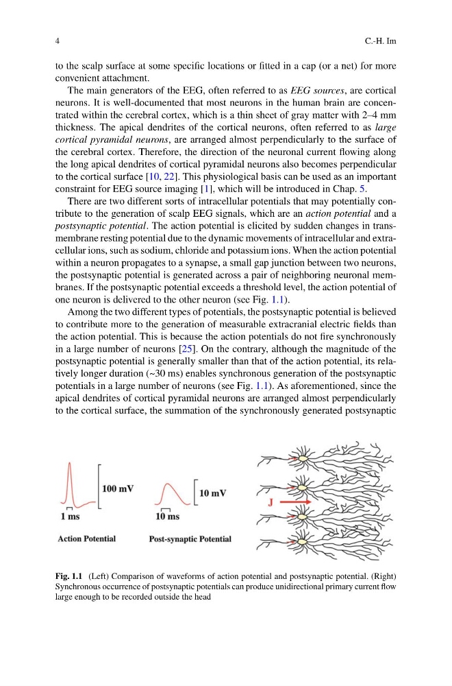
There are two different sorts of intracellular potentials that may potentially contribute to the generation of scalp EEG signals, which are an action potential and a
postsynaptic potential. The action potential is elicited by sudden changes in transmembrane resting potential due to the dynamic movements of intracellular and extracellular ions, such as sodium, chloride and potassium ions. When the action potential
within a neuron propagates to a synapse, a small gap junction between two neurons,
the postsynaptic potential is generated across a pair of neighboring neuronal membranes. If the postsynaptic potential exceeds a threshold level, the action potential of
one neuron is delivered to the other neuron (see Fig. 1.1).
Among the two different types of potentials, the postsynaptic potential is believed
to contribute more to the generation of measurable extracranial electric fields than
the action potential. This is because the action potentials do not fire synchronously
in a large number of neurons [25]. On the contrary, although the magnitude of the
postsynaptic potential is generally smaller than that of the action potential, its relatively longer duration (~30 ms) enables synchronous generation of the postsynaptic
potentials in a large number of neurons (see Fig. 1.1). As aforementioned, since the
apical dendrites of cortical pyramidal neurons are arranged almost perpendicularly
to the cortical surface, the summation of the synchronously generated postsynaptic
Fig. 1.1 (Left) Comparison of waveforms of action potential and postsynaptic potential. (Right)
Synchronous occurrence of postsynaptic potentials can produce unidirectional primary current flow
large enough to be recorded outside the head
C.-H. Im
to the scalp surface at some specific locations or fitted in a cap (or a net) for more
convenient attachment.
The main generators of the EEG, often referred to as EEG sources, are cortical
neurons. It is well-documented that most neurons in the human brain are concentrated within the cerebral cortex, which is a thin sheet of gray matter with 2–4 mm
thickness. The apical dendrites of the cortical neurons, often referred to as large
cortical pyramidal neurons, are arranged almost perpendicularly to the surface of
the cerebral cortex. Therefore, the direction of the neuronal current flowing along
the long apical dendrites of cortical pyramidal neurons also becomes perpendicular
to the cortical surface [10, 22]. This physiological basis can be used as an important
constraint for EEG source imaging [1], which will be introduced in Chap. 5.
There are two different sorts of intracellular potentials that may potentially contribute to the generation of scalp EEG signals, which are an action potential and a
postsynaptic potential. The action potential is elicited by sudden changes in transmembrane resting potential due to the dynamic movements of intracellular and extracellular ions, such as sodium, chloride and potassium ions. When the action potential
within a neuron propagates to a synapse, a small gap junction between two neurons,
the postsynaptic potential is generated across a pair of neighboring neuronal membranes. If the postsynaptic potential exceeds a threshold level, the action potential of
one neuron is delivered to the other neuron (see Fig. 1.1).
Among the two different types of potentials, the postsynaptic potential is believed
to contribute more to the generation of measurable extracranial electric fields than
the action potential. This is because the action potentials do not fire synchronously
in a large number of neurons [25]. On the contrary, although the magnitude of the
postsynaptic potential is generally smaller than that of the action potential, its relatively longer duration (~30 ms) enables synchronous generation of the postsynaptic
potentials in a large number of neurons (see Fig. 1.1). As aforementioned, since the
apical dendrites of cortical pyramidal neurons are arranged almost perpendicularly
to the cortical surface, the summation of the synchronously generated postsynaptic
Fig. 1.1 (Left) Comparison of waveforms of action potential and postsynaptic potential. (Right)
Synchronous occurrence of postsynaptic potentials can produce unidirectional primary current flow
large enough to be recorded outside the head