

118
Y. Zhang
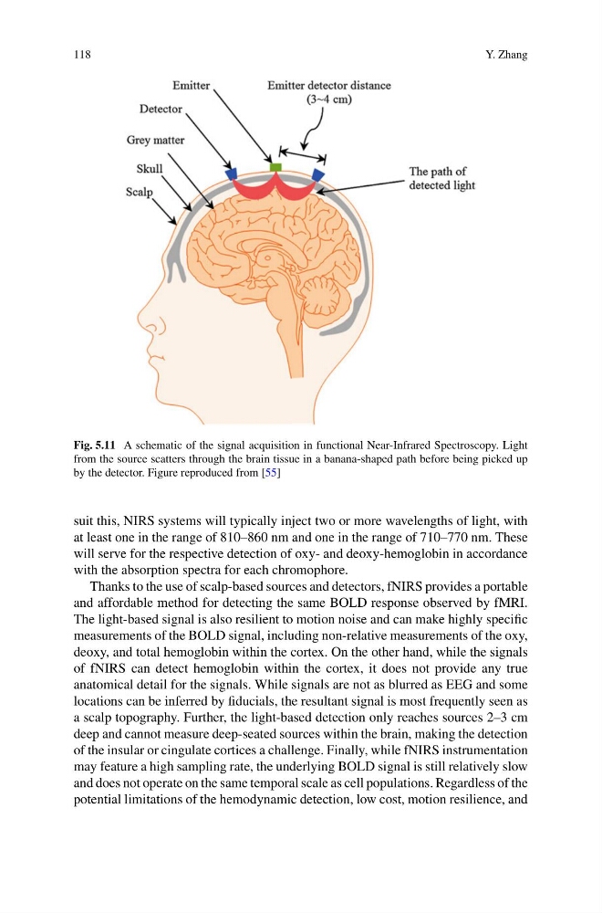
Fig. 5.11 A schematic of the signal acquisition in functional Near-Infrared Spectroscopy. Light
from the source scatters through the brain tissue in a banana-shaped path before being picked up
by the detector. Figure reproduced from [55]
suit this, NIRS systems will typically inject two or more wavelengths of light, with
at least one in the range of 810–860 nm and one in the range of 710–770 nm. These
will serve for the respective detection of oxy- and deoxy-hemoglobin in accordance
with the absorption spectra for each chromophore.
Thanks to the use of scalp-based sources and detectors, fNIRS provides a portable
and affordable method for detecting the same BOLD response observed by fMRI.
The light-based signal is also resilient to motion noise and can make highly specific
measurements of the BOLD signal, including non-relative measurements of the oxy,
deoxy, and total hemoglobin within the cortex. On the other hand, while the signals
of fNIRS can detect hemoglobin within the cortex, it does not provide any true
anatomical detail for the signals. While signals are not as blurred as EEG and some
locations can be inferred by fiducials, the resultant signal is most frequently seen as
a scalp topography. Further, the light-based detection only reaches sources 2–3 cm
deep and cannot measure deep-seated sources within the brain, making the detection
of the insular or cingulate cortices a challenge. Finally, while fNIRS instrumentation
may feature a high sampling rate, the underlying BOLD signal is still relatively slow
and does not operate on the same temporal scale as cell populations. Regardless of the
potential limitations of the hemodynamic detection, low cost, motion resilience, and
Y. Zhang
Fig. 5.11 A schematic of the signal acquisition in functional Near-Infrared Spectroscopy. Light
from the source scatters through the brain tissue in a banana-shaped path before being picked up
by the detector. Figure reproduced from [55]
suit this, NIRS systems will typically inject two or more wavelengths of light, with
at least one in the range of 810–860 nm and one in the range of 710–770 nm. These
will serve for the respective detection of oxy- and deoxy-hemoglobin in accordance
with the absorption spectra for each chromophore.
Thanks to the use of scalp-based sources and detectors, fNIRS provides a portable
and affordable method for detecting the same BOLD response observed by fMRI.
The light-based signal is also resilient to motion noise and can make highly specific
measurements of the BOLD signal, including non-relative measurements of the oxy,
deoxy, and total hemoglobin within the cortex. On the other hand, while the signals
of fNIRS can detect hemoglobin within the cortex, it does not provide any true
anatomical detail for the signals. While signals are not as blurred as EEG and some
locations can be inferred by fiducials, the resultant signal is most frequently seen as
a scalp topography. Further, the light-based detection only reaches sources 2–3 cm
deep and cannot measure deep-seated sources within the brain, making the detection
of the insular or cingulate cortices a challenge. Finally, while fNIRS instrumentation
may feature a high sampling rate, the underlying BOLD signal is still relatively slow
and does not operate on the same temporal scale as cell populations. Regardless of the
potential limitations of the hemodynamic detection, low cost, motion resilience, and