

years, attributed possibly to the potential toxicity of carbon nanomaterials over
long-term. Nonetheless the development of CNT-based imaging agents still has
room to grow when it comes to multimodality nuclear imaging applications [136].
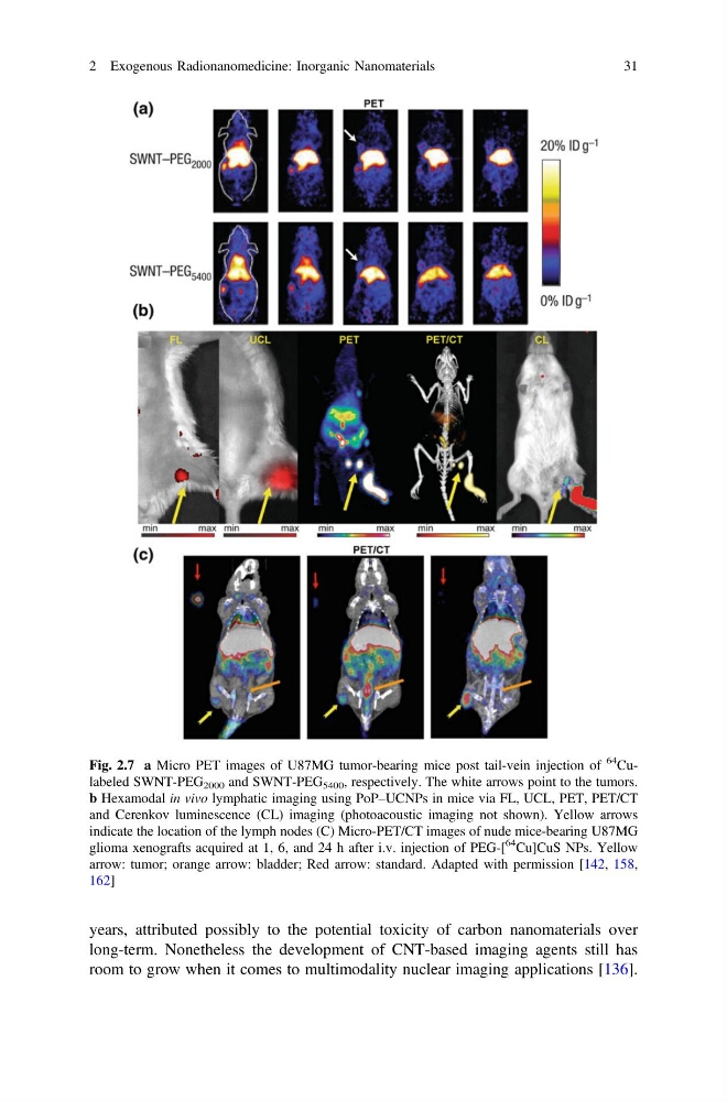
Fig. 2.7 a Micro PET images of U87MG tumor-bearing mice post tail-vein injection of
64
Culabeled SWNT-PEG 2000 and SWNT-PEG 5400 , respectively. The white arrows point to the tumors.
b Hexamodal in vivo lymphatic imaging using PoP–UCNPs in mice via FL, UCL, PET, PET/CT
and Cerenkov luminescence (CL) imaging (photoacoustic imaging not shown). Yellow arrows
indicate the location of the lymph nodes (C) Micro-PET/CT images of nude mice-bearing U87MG
glioma xenografts acquired at 1, 6, and 24 h after i.v. injection of PEG-[
64
Cu]CuS NPs. Yellow
arrow: tumor; orange arrow: bladder; Red arrow: standard. Adapted with permission [142, 158,
162]
2 Exogenous Radionanomedicine: Inorganic Nanomaterials
31
long-term. Nonetheless the development of CNT-based imaging agents still has
room to grow when it comes to multimodality nuclear imaging applications [136].
Fig. 2.7 a Micro PET images of U87MG tumor-bearing mice post tail-vein injection of
64
Culabeled SWNT-PEG 2000 and SWNT-PEG 5400 , respectively. The white arrows point to the tumors.
b Hexamodal in vivo lymphatic imaging using PoP–UCNPs in mice via FL, UCL, PET, PET/CT
and Cerenkov luminescence (CL) imaging (photoacoustic imaging not shown). Yellow arrows
indicate the location of the lymph nodes (C) Micro-PET/CT images of nude mice-bearing U87MG
glioma xenografts acquired at 1, 6, and 24 h after i.v. injection of PEG-[
64
Cu]CuS NPs. Yellow
arrow: tumor; orange arrow: bladder; Red arrow: standard. Adapted with permission [142, 158,
162]
2 Exogenous Radionanomedicine: Inorganic Nanomaterials
31