

a fast, greater renal excretion, and shorter blood circulation half-life. No radioactive
tag was conjugated to these particles, and all of estimations were purely based on
optical imaging and quantification in this research.
To better quantify the PK profile of silica nanoparticles and ensure a specific
tumor active targeting in vivo, two years later, the same group reported the systematic pre-clinical study in both small and large animals by further upgrading their
platform with cRGDY (small peptides that target integrin a v b 3 from human melanoma) and radioiodine (
124 I, t 1/2 = 4.2 days), forming the first generation of
tumor-targeting C dots [35]. The final HD size was well-controlled to be around
7 nm with about 6–7 cRGDY ligands in each C dot. Systematic in vitro
receptor-binding studies showed high and specific binding affinity of
124 I–cRGDY–
PEG–C dots. In vivo tumor targeting, biodistribution and clearance studies were
then performed in M21 tumor xenograft models. When compared with small
peptides, which usually have less than 10 min blood circulation half-life,
124 I–
cRGDY–PEG–C dots showed a nearly 6 h half-life in vivo. Efficient renal excretion was found in both targeted and non-targeted groups, with nearly 50%ID
excreted within 24 h and over 70%ID by 96 h. Whole body PET imaging revealed
a 3-fold higher nanoparticle uptake in M21 tumors in the targeted group than the
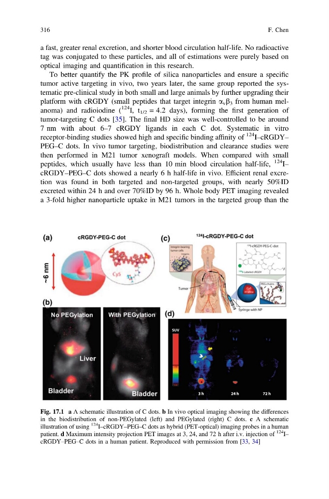
Fig. 17.1 a A schematic illustration of C dots. b In vivo optical imaging showing the differences
in the biodistribution of non-PEGylated (left) and PEGylated (right) C dots. c A schematic
illustration of using
124
I–cRGDY–PEG–C dots as hybrid (PET-optical) imaging probes in a human
patient. d Maximum intensity projection PET images at 3, 24, and 72 h after i.v. injection of
124
I–
cRGDY–PEG–C dots in a human patient. Reproduced with permission from [33, 34]
316
F. Chen
tag was conjugated to these particles, and all of estimations were purely based on
optical imaging and quantification in this research.
To better quantify the PK profile of silica nanoparticles and ensure a specific
tumor active targeting in vivo, two years later, the same group reported the systematic pre-clinical study in both small and large animals by further upgrading their
platform with cRGDY (small peptides that target integrin a v b 3 from human melanoma) and radioiodine (
124 I, t 1/2 = 4.2 days), forming the first generation of
tumor-targeting C dots [35]. The final HD size was well-controlled to be around
7 nm with about 6–7 cRGDY ligands in each C dot. Systematic in vitro
receptor-binding studies showed high and specific binding affinity of
124 I–cRGDY–
PEG–C dots. In vivo tumor targeting, biodistribution and clearance studies were
then performed in M21 tumor xenograft models. When compared with small
peptides, which usually have less than 10 min blood circulation half-life,
124 I–
cRGDY–PEG–C dots showed a nearly 6 h half-life in vivo. Efficient renal excretion was found in both targeted and non-targeted groups, with nearly 50%ID
excreted within 24 h and over 70%ID by 96 h. Whole body PET imaging revealed
a 3-fold higher nanoparticle uptake in M21 tumors in the targeted group than the
Fig. 17.1 a A schematic illustration of C dots. b In vivo optical imaging showing the differences
in the biodistribution of non-PEGylated (left) and PEGylated (right) C dots. c A schematic
illustration of using
124
I–cRGDY–PEG–C dots as hybrid (PET-optical) imaging probes in a human
patient. d Maximum intensity projection PET images at 3, 24, and 72 h after i.v. injection of
124
I–
cRGDY–PEG–C dots in a human patient. Reproduced with permission from [33, 34]
316
F. Chen