

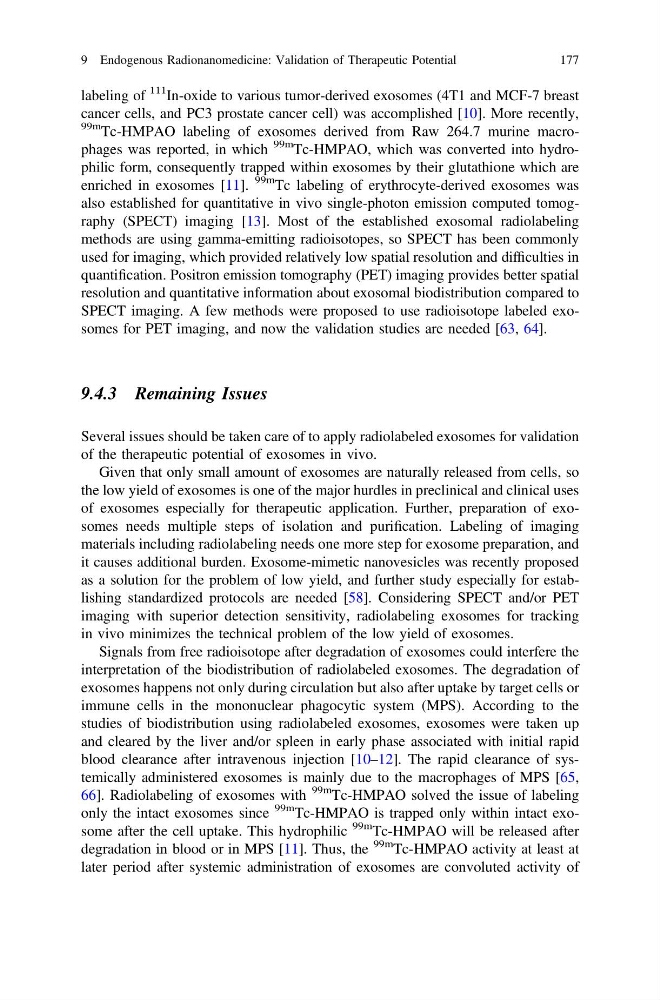
labeling of
111 In-oxide to various tumor-derived exosomes (4T1 and MCF-7 breast
cancer cells, and PC3 prostate cancer cell) was accomplished [10]. More recently,
99m Tc-HMPAO labeling of exosomes derived from Raw 264.7 murine macrophages was reported, in which
99m
Tc-HMPAO, which was converted into hydrophilic form, consequently trapped within exosomes by their glutathione which are
enriched in exosomes [11].
99m Tc labeling of erythrocyte-derived exosomes was
also established for quantitative in vivo single-photon emission computed tomography (SPECT) imaging [13]. Most of the established exosomal radiolabeling
methods are using gamma-emitting radioisotopes, so SPECT has been commonly
used for imaging, which provided relatively low spatial resolution and difficulties in
quantification. Positron emission tomography (PET) imaging provides better spatial
resolution and quantitative information about exosomal biodistribution compared to
SPECT imaging. A few methods were proposed to use radioisotope labeled exosomes for PET imaging, and now the validation studies are needed [63, 64].
9.4.3 Remaining Issues
Several issues should be taken care of to apply radiolabeled exosomes for validation
of the therapeutic potential of exosomes in vivo.
Given that only small amount of exosomes are naturally released from cells, so
the low yield of exosomes is one of the major hurdles in preclinical and clinical uses
of exosomes especially for therapeutic application. Further, preparation of exosomes needs multiple steps of isolation and purification. Labeling of imaging
materials including radiolabeling needs one more step for exosome preparation, and
it causes additional burden. Exosome-mimetic nanovesicles was recently proposed
as a solution for the problem of low yield, and further study especially for establishing standardized protocols are needed [58]. Considering SPECT and/or PET
imaging with superior detection sensitivity, radiolabeling exosomes for tracking
in vivo minimizes the technical problem of the low yield of exosomes.
Signals from free radioisotope after degradation of exosomes could interfere the
interpretation of the biodistribution of radiolabeled exosomes. The degradation of
exosomes happens not only during circulation but also after uptake by target cells or
immune cells in the mononuclear phagocytic system (MPS). According to the
studies of biodistribution using radiolabeled exosomes, exosomes were taken up
and cleared by the liver and/or spleen in early phase associated with initial rapid
blood clearance after intravenous injection [10–12]. The rapid clearance of systemically administered exosomes is mainly due to the macrophages of MPS [65,
66]. Radiolabeling of exosomes with
99m Tc-HMPAO solved the issue of labeling
only the intact exosomes since
99m Tc-HMPAO is trapped only within intact exosome after the cell uptake. This hydrophilic
99m Tc-HMPAO will be released after
degradation in blood or in MPS [11]. Thus, the
99m
Tc-HMPAO activity at least at
later period after systemic administration of exosomes are convoluted activity of
9 Endogenous Radionanomedicine: Validation of Therapeutic Potential
177
111 In-oxide to various tumor-derived exosomes (4T1 and MCF-7 breast
cancer cells, and PC3 prostate cancer cell) was accomplished [10]. More recently,
99m Tc-HMPAO labeling of exosomes derived from Raw 264.7 murine macrophages was reported, in which
99m
Tc-HMPAO, which was converted into hydrophilic form, consequently trapped within exosomes by their glutathione which are
enriched in exosomes [11].
99m Tc labeling of erythrocyte-derived exosomes was
also established for quantitative in vivo single-photon emission computed tomography (SPECT) imaging [13]. Most of the established exosomal radiolabeling
methods are using gamma-emitting radioisotopes, so SPECT has been commonly
used for imaging, which provided relatively low spatial resolution and difficulties in
quantification. Positron emission tomography (PET) imaging provides better spatial
resolution and quantitative information about exosomal biodistribution compared to
SPECT imaging. A few methods were proposed to use radioisotope labeled exosomes for PET imaging, and now the validation studies are needed [63, 64].
9.4.3 Remaining Issues
Several issues should be taken care of to apply radiolabeled exosomes for validation
of the therapeutic potential of exosomes in vivo.
Given that only small amount of exosomes are naturally released from cells, so
the low yield of exosomes is one of the major hurdles in preclinical and clinical uses
of exosomes especially for therapeutic application. Further, preparation of exosomes needs multiple steps of isolation and purification. Labeling of imaging
materials including radiolabeling needs one more step for exosome preparation, and
it causes additional burden. Exosome-mimetic nanovesicles was recently proposed
as a solution for the problem of low yield, and further study especially for establishing standardized protocols are needed [58]. Considering SPECT and/or PET
imaging with superior detection sensitivity, radiolabeling exosomes for tracking
in vivo minimizes the technical problem of the low yield of exosomes.
Signals from free radioisotope after degradation of exosomes could interfere the
interpretation of the biodistribution of radiolabeled exosomes. The degradation of
exosomes happens not only during circulation but also after uptake by target cells or
immune cells in the mononuclear phagocytic system (MPS). According to the
studies of biodistribution using radiolabeled exosomes, exosomes were taken up
and cleared by the liver and/or spleen in early phase associated with initial rapid
blood clearance after intravenous injection [10–12]. The rapid clearance of systemically administered exosomes is mainly due to the macrophages of MPS [65,
66]. Radiolabeling of exosomes with
99m Tc-HMPAO solved the issue of labeling
only the intact exosomes since
99m Tc-HMPAO is trapped only within intact exosome after the cell uptake. This hydrophilic
99m Tc-HMPAO will be released after
degradation in blood or in MPS [11]. Thus, the
99m
Tc-HMPAO activity at least at
later period after systemic administration of exosomes are convoluted activity of
9 Endogenous Radionanomedicine: Validation of Therapeutic Potential
177