

After two years, Shi et al. demonstrated the use of iron oxide nanoparticle
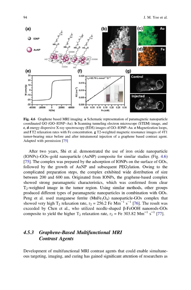
(IONPs)–GOs–gold nanoparticle (AuNP) composite for similar studies (Fig. 4.6)
[75]. The complex was prepared by the adsorption of IONPs on the surface of GOs,
followed by the growth of AuNP and subsequent PEGylation. Owing to the
complicated preparation steps, the complex exhibited wide distribution of size
between 200 and 600 nm. Originated from IONPs, the graphene-based complex
showed strong paramagnetic characteristics, which was confirmed from clear
T 2 -weighted image in the tumor region. Using similar methods, other groups
produced different types of paramagnetic nanoparticles in combination with GOs.
Peng et al. used manganese ferrite (MnFe 2 O 4 ) nanoparticle-GOs complex that
showed very high T 2 relaxation rate, r 2 = 256.2 Fe Mm
−1 s
−1 [76]. The result was
exceeded by Chen et al., who utilized needle-shaped b-FeOOH nanorods-GOs
composite to yield the higher T 2 relaxation rate, r 2 = Fe 303.82 Mm
−1 s
−1 [77].
4.5.3 Graphene-Based Multifunctional MRI
Contrast Agents
Development of multifunctional MRI contrast agents that could enable simultaneous targeting, imaging, and curing has gained significant attention of researchers as
Fig. 4.6 Graphene based MRI imaging. a Schematic representation of paramagnetic nanoparticle
coordinated GO (GO–IONP–Au). b Scanning tunneling electron microscope (STEM) image, and
c, d energy dispersive X-ray spectroscopy (EDS) images of GO–IONP–Au. e Magnetization loops,
and f T2 relaxation rates with Fe concentration. g T2-weighted magnetic resonance images of 4T1
tumor-bearing mice before and after intratumoral injection of a graphene based contrast agent.
Adapted with permission [75]
94
J. M. Yoo et al.
(IONPs)–GOs–gold nanoparticle (AuNP) composite for similar studies (Fig. 4.6)
[75]. The complex was prepared by the adsorption of IONPs on the surface of GOs,
followed by the growth of AuNP and subsequent PEGylation. Owing to the
complicated preparation steps, the complex exhibited wide distribution of size
between 200 and 600 nm. Originated from IONPs, the graphene-based complex
showed strong paramagnetic characteristics, which was confirmed from clear
T 2 -weighted image in the tumor region. Using similar methods, other groups
produced different types of paramagnetic nanoparticles in combination with GOs.
Peng et al. used manganese ferrite (MnFe 2 O 4 ) nanoparticle-GOs complex that
showed very high T 2 relaxation rate, r 2 = 256.2 Fe Mm
−1 s
−1 [76]. The result was
exceeded by Chen et al., who utilized needle-shaped b-FeOOH nanorods-GOs
composite to yield the higher T 2 relaxation rate, r 2 = Fe 303.82 Mm
−1 s
−1 [77].
4.5.3 Graphene-Based Multifunctional MRI
Contrast Agents
Development of multifunctional MRI contrast agents that could enable simultaneous targeting, imaging, and curing has gained significant attention of researchers as
Fig. 4.6 Graphene based MRI imaging. a Schematic representation of paramagnetic nanoparticle
coordinated GO (GO–IONP–Au). b Scanning tunneling electron microscope (STEM) image, and
c, d energy dispersive X-ray spectroscopy (EDS) images of GO–IONP–Au. e Magnetization loops,
and f T2 relaxation rates with Fe concentration. g T2-weighted magnetic resonance images of 4T1
tumor-bearing mice before and after intratumoral injection of a graphene based contrast agent.
Adapted with permission [75]
94
J. M. Yoo et al.