

4.5 Recent Applications of Graphene-Based
Nanomaterials in Magnetic Resonance Imaging
MRI is the most powerful technique for imaging tumors, the cardiac and nervous
systems as it enables high-resolution imaging in a non-invasive manner [67].
However, when a diseased lesion do not show distinct change in relaxation times,
MRI imaging does not help discover the diseased areas. MRI contrast agents are
employed to maximize the difference in relaxation time. Currently, most of the
commercial contrast agents are paramagnetic nanoparticles and metal ion complexes [68]. Graphene-based nanomaterials do not exhibit intrinsic paramagnetism
and is not a candidate as a novel MRI contrast agent. However, functionalized
graphene-based nanomaterial possess oxygenated functional groups and cavities
that can load various drugs with conventional MRI contrast agents. In addition,
appropriate edge modifications can enable efficient targeting to specific regions.
Moreover, docking MRI contrast agents inside graphene-based nanomaterials can
also alleviate the toxicity concerns of heavy metal ions by decreasing the release
rate.
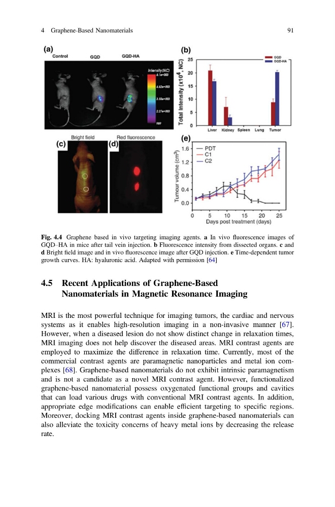
Fig. 4.4 Graphene based in vivo targeting imaging agents. a In vivo fluorescence images of
GQD–HA in mice after tail vein injection. b Fluorescence intensity from dissected organs. c and
d Bright field image and in vivo fluorescence image after GQD injection. e Time-dependent tumor
growth curves. HA: hyaluronic acid. Adapted with permission [64]
4 Graphene-Based Nanomaterials
91
Nanomaterials in Magnetic Resonance Imaging
MRI is the most powerful technique for imaging tumors, the cardiac and nervous
systems as it enables high-resolution imaging in a non-invasive manner [67].
However, when a diseased lesion do not show distinct change in relaxation times,
MRI imaging does not help discover the diseased areas. MRI contrast agents are
employed to maximize the difference in relaxation time. Currently, most of the
commercial contrast agents are paramagnetic nanoparticles and metal ion complexes [68]. Graphene-based nanomaterials do not exhibit intrinsic paramagnetism
and is not a candidate as a novel MRI contrast agent. However, functionalized
graphene-based nanomaterial possess oxygenated functional groups and cavities
that can load various drugs with conventional MRI contrast agents. In addition,
appropriate edge modifications can enable efficient targeting to specific regions.
Moreover, docking MRI contrast agents inside graphene-based nanomaterials can
also alleviate the toxicity concerns of heavy metal ions by decreasing the release
rate.
Fig. 4.4 Graphene based in vivo targeting imaging agents. a In vivo fluorescence images of
GQD–HA in mice after tail vein injection. b Fluorescence intensity from dissected organs. c and
d Bright field image and in vivo fluorescence image after GQD injection. e Time-dependent tumor
growth curves. HA: hyaluronic acid. Adapted with permission [64]
4 Graphene-Based Nanomaterials
91