

4.3.3 Biodegradation
In addition to various in vitro/in vivo toxicity studies, some researches devoted
special attention to potential oxidative biodegradation of graphene-based nanomaterials by intracellular enzymes. In 2011 and 2012, Kotchey et al. reported two
studies on enzyme-catalyzed degradation of graphene-based nanomaterials.
Foremost, the authors utilized low concentrations of horseradish peroxidase
(HRP) to verify their effects on GOs and rGOs [47]. Obtained atomic force
microscopy (AFM) and transmission electron microscopy images as well as Raman
spectra concordantly showed that HRP caused the generation of holey GOs, which
resulted in debris of oxidized GO flakes in the end. Contrarily, HRP did not induce
any structural changes on rGOs, which implied that the presence of functional
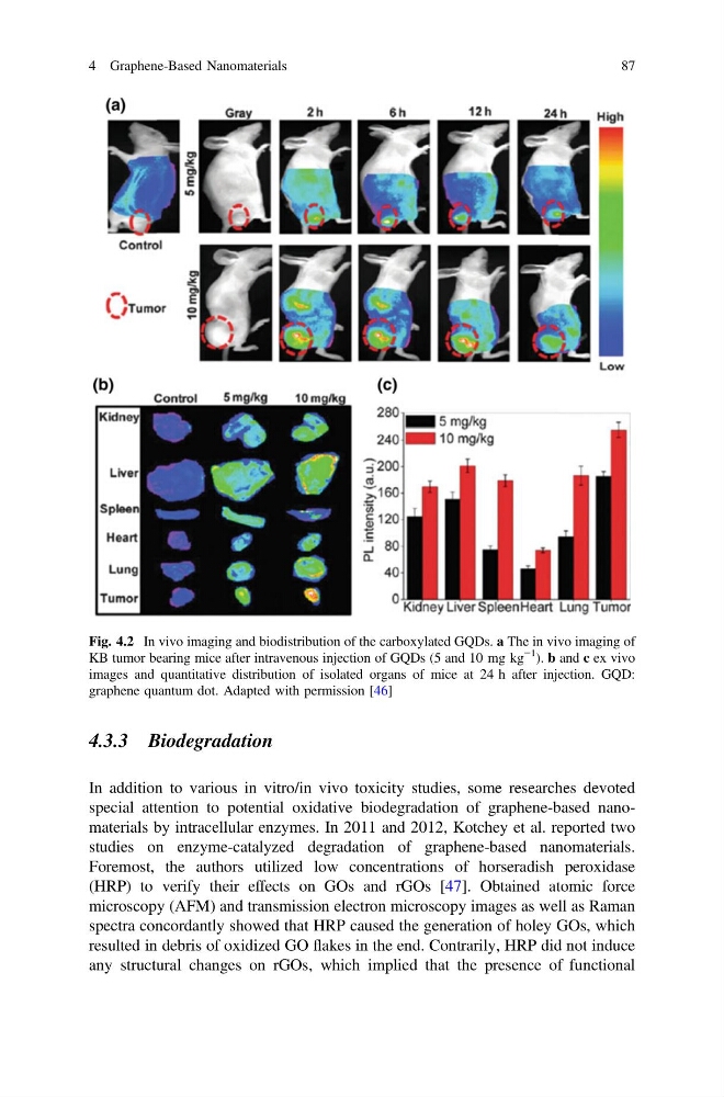
Fig. 4.2 In vivo imaging and biodistribution of the carboxylated GQDs. a The in vivo imaging of
KB tumor bearing mice after intravenous injection of GQDs (5 and 10 mg kg
−1
). b and c ex vivo
images and quantitative distribution of isolated organs of mice at 24 h after injection. GQD:
graphene quantum dot. Adapted with permission [46]
4 Graphene-Based Nanomaterials
87
In addition to various in vitro/in vivo toxicity studies, some researches devoted
special attention to potential oxidative biodegradation of graphene-based nanomaterials by intracellular enzymes. In 2011 and 2012, Kotchey et al. reported two
studies on enzyme-catalyzed degradation of graphene-based nanomaterials.
Foremost, the authors utilized low concentrations of horseradish peroxidase
(HRP) to verify their effects on GOs and rGOs [47]. Obtained atomic force
microscopy (AFM) and transmission electron microscopy images as well as Raman
spectra concordantly showed that HRP caused the generation of holey GOs, which
resulted in debris of oxidized GO flakes in the end. Contrarily, HRP did not induce
any structural changes on rGOs, which implied that the presence of functional
Fig. 4.2 In vivo imaging and biodistribution of the carboxylated GQDs. a The in vivo imaging of
KB tumor bearing mice after intravenous injection of GQDs (5 and 10 mg kg
−1
). b and c ex vivo
images and quantitative distribution of isolated organs of mice at 24 h after injection. GQD:
graphene quantum dot. Adapted with permission [46]
4 Graphene-Based Nanomaterials
87