

frequently employed for biomedical applications while the term graphene refers to a
two-dimensional atomic film. GOs and GQDs are especially broadly utilized for
biomedical applications due to their high dispersibility and low cytotoxicity.
Briefly, GOs are synthesized through the Hummer’s method and GQDs can be
obtained by thermo-oxidative cutting of carbon precursors including GOs. Notably,
GOs and GQDs have various functional groups available for further modifications,
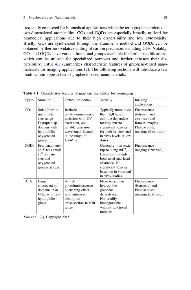
which can be utilized for specialized purposes and further enhance their dispersibility. Table 4.1 summarizes characteristic features of graphene-based nanomaterials for imaging applications [2]. The following sections will introduce a few
modification approaches of graphene-based nanomaterials.
Table 4.1 Characteristic features of graphene derivatives for bioimaging
Types Structure
Optical properties
Toxicity
Imaging
applications
GOs
Sub-10 nm to
micrometer
size range
Disrupted sp
2
domain with
hydrophilic
oxygenated
group
Intrinsic
photo-luminescence
emission with UV
excitation, and
tunable emission
wavelength located
at the range of
UV–Vis
Typically more toxic
than GQDs, and
cell-line dependent
toxicity but no
significant toxicity
for both in vitro and
in vivo levels at low
doses
Fluorescence
(Intrinsic and
extrinsic) and
Raman imaging,
Photoacoustic
imaging (Extrinsic)
GQDs Few nanometer
(2–5 nm) small
sp
2 domain
size and
oxygenated
groups at edge
Generally, non-toxic
(up to 1 mg ml
−1
).
Excretion through
both renal and fecal
clearance. No
significant toxicity
based on in vitro and
in vivo studies
Fluorescence
imaging (Intrinsic)
rGOs Large
connected sp
2
domains than
GOs, with few
hydrophilic
group
A high
photoluminescence
quenching effect
with enhanced
absorption
cross-section in NIR
range
More toxic than
hydrophilic
graphene
derivatives.
Not-readily
biodegradable
without functional
moieties
Fluorescence
(Extrinsic) and
Photoacoustic
imaging (Intrinsic)
Yoo et al. [2], Copyright 2015
4 Graphene-Based Nanomaterials
81
two-dimensional atomic film. GOs and GQDs are especially broadly utilized for
biomedical applications due to their high dispersibility and low cytotoxicity.
Briefly, GOs are synthesized through the Hummer’s method and GQDs can be
obtained by thermo-oxidative cutting of carbon precursors including GOs. Notably,
GOs and GQDs have various functional groups available for further modifications,
which can be utilized for specialized purposes and further enhance their dispersibility. Table 4.1 summarizes characteristic features of graphene-based nanomaterials for imaging applications [2]. The following sections will introduce a few
modification approaches of graphene-based nanomaterials.
Table 4.1 Characteristic features of graphene derivatives for bioimaging
Types Structure
Optical properties
Toxicity
Imaging
applications
GOs
Sub-10 nm to
micrometer
size range
Disrupted sp
2
domain with
hydrophilic
oxygenated
group
Intrinsic
photo-luminescence
emission with UV
excitation, and
tunable emission
wavelength located
at the range of
UV–Vis
Typically more toxic
than GQDs, and
cell-line dependent
toxicity but no
significant toxicity
for both in vitro and
in vivo levels at low
doses
Fluorescence
(Intrinsic and
extrinsic) and
Raman imaging,
Photoacoustic
imaging (Extrinsic)
GQDs Few nanometer
(2–5 nm) small
sp
2 domain
size and
oxygenated
groups at edge
Generally, non-toxic
(up to 1 mg ml
−1
).
Excretion through
both renal and fecal
clearance. No
significant toxicity
based on in vitro and
in vivo studies
Fluorescence
imaging (Intrinsic)
rGOs Large
connected sp
2
domains than
GOs, with few
hydrophilic
group
A high
photoluminescence
quenching effect
with enhanced
absorption
cross-section in NIR
range
More toxic than
hydrophilic
graphene
derivatives.
Not-readily
biodegradable
without functional
moieties
Fluorescence
(Extrinsic) and
Photoacoustic
imaging (Intrinsic)
Yoo et al. [2], Copyright 2015
4 Graphene-Based Nanomaterials
81