

a motif homologous to the NAC domain (PF02365) (Pascual et al. 2015), which is
present in some NAC proteins (BpNAC4 and BpNAC12) from Betula platyphylla
(Guo et al. 2017) and in a small number of NAC proteins from other plants (Hussey
et al. 2015). In other species, a premature stop codon after the NAC domain has been
identified, while other NAC proteins present NAC domain repeats in tandem (Singh
et al. 2013). Although some studies suggest that the typical structure of the five subdomains (A–E) or at least subdomains C and D are essential for the accurate functioning of the NAC TFs, additional research is needed to elucidate the role of these
structural modifications in some family members.
Despite the numerous studies and scientific interest in the NAC family, only two
crystal structures of NAC domains have been reported. One structure corresponds to
the NAC domain from the Arabidopsis ANAC019 protein (Ernst et al. 2004), and the
other corresponds to the rice SNAC1 protein (Chen et al. 2011). The NAC domain of
ANAC019 shares structural similarity with AtNAC1 (Zhu et al. 2014) and StNAC
from Solanum tuberosum (Singh et al. 2013) and BdNAC from Brachypodium
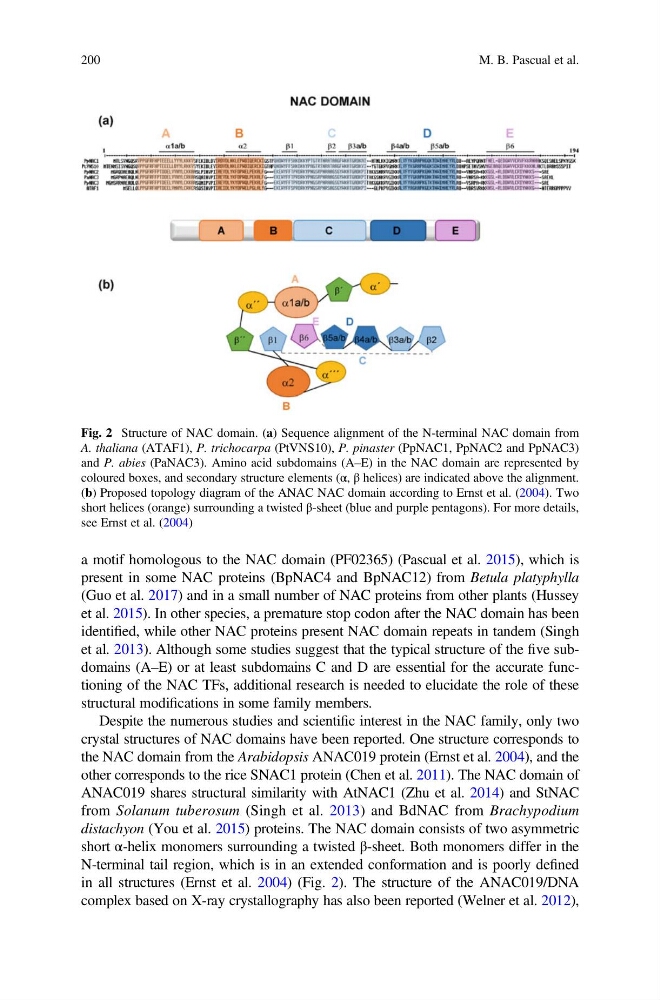
distachyon (You et al. 2015) proteins. The NAC domain consists of two asymmetric
short α-helix monomers surrounding a twisted β-sheet. Both monomers differ in the
N-terminal tail region, which is in an extended conformation and is poorly defined
in all structures (Ernst et al. 2004) (Fig. 2). The structure of the ANAC019/DNA
complex based on X-ray crystallography has also been reported (Welner et al. 2012),
Fig. 2 Structure of NAC domain. (a) Sequence alignment of the N-terminal NAC domain from
A. thaliana (ATAF1), P. trichocarpa (PtVNS10), P. pinaster (PpNAC1, PpNAC2 and PpNAC3)
and P. abies (PaNAC3). Amino acid subdomains (A–E) in the NAC domain are represented by
coloured boxes, and secondary structure elements (α, β helices) are indicated above the alignment.
(b) Proposed topology diagram of the ANAC NAC domain according to Ernst et al. (2004). Two
short helices (orange) surrounding a twisted β-sheet (blue and purple pentagons). For more details,
see Ernst et al. (2004)
200
M. B. Pascual et al.
present in some NAC proteins (BpNAC4 and BpNAC12) from Betula platyphylla
(Guo et al. 2017) and in a small number of NAC proteins from other plants (Hussey
et al. 2015). In other species, a premature stop codon after the NAC domain has been
identified, while other NAC proteins present NAC domain repeats in tandem (Singh
et al. 2013). Although some studies suggest that the typical structure of the five subdomains (A–E) or at least subdomains C and D are essential for the accurate functioning of the NAC TFs, additional research is needed to elucidate the role of these
structural modifications in some family members.
Despite the numerous studies and scientific interest in the NAC family, only two
crystal structures of NAC domains have been reported. One structure corresponds to
the NAC domain from the Arabidopsis ANAC019 protein (Ernst et al. 2004), and the
other corresponds to the rice SNAC1 protein (Chen et al. 2011). The NAC domain of
ANAC019 shares structural similarity with AtNAC1 (Zhu et al. 2014) and StNAC
from Solanum tuberosum (Singh et al. 2013) and BdNAC from Brachypodium
distachyon (You et al. 2015) proteins. The NAC domain consists of two asymmetric
short α-helix monomers surrounding a twisted β-sheet. Both monomers differ in the
N-terminal tail region, which is in an extended conformation and is poorly defined
in all structures (Ernst et al. 2004) (Fig. 2). The structure of the ANAC019/DNA
complex based on X-ray crystallography has also been reported (Welner et al. 2012),
Fig. 2 Structure of NAC domain. (a) Sequence alignment of the N-terminal NAC domain from
A. thaliana (ATAF1), P. trichocarpa (PtVNS10), P. pinaster (PpNAC1, PpNAC2 and PpNAC3)
and P. abies (PaNAC3). Amino acid subdomains (A–E) in the NAC domain are represented by
coloured boxes, and secondary structure elements (α, β helices) are indicated above the alignment.
(b) Proposed topology diagram of the ANAC NAC domain according to Ernst et al. (2004). Two
short helices (orange) surrounding a twisted β-sheet (blue and purple pentagons). For more details,
see Ernst et al. (2004)
200
M. B. Pascual et al.