

would expect by chance, it is likely that these sequences are found near each other in
the nucleus and are likely parts of the same chromosome. It was through Hi-C
research that topographically associating domains (TADs) were discovered. Hi-C
can be used to gain insight into sequences involved in long-distance (along the linear
DNA molecule) interactions (e.g., promoters and enhancer elements) (Belton et al.
2012; Denker and De Laat 2016).
Of note, Hi-C can be a form of phasing. Homologous chromosomes occupy their
own regions within the 3D-space of a nucleus. Thus formaldehyde cross-linking
between chromatin from homologous chromosomes is unlikely. If Hi-C sequencing
d
c
b
a
h
g
f
e
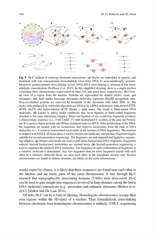
Fig. 9 Hi-C method of studying chromatin interactions. (a) Nuclei are embedded in agarose, and
treatment with low concentration formaldehyde cross-links DNA to associated/nearby proteins.
Moreover, protein-protein cross-linking occurs. DNA-DNA cross-linking is minimal at low formaldehyde concentration (Hoffman et al. 2015). In this simplified drawing, there is a single nucleus
containing three chromosomes (represented by blue, red, and green lines, respectively). (b) Closeup view of a region from the nucleus. Proteins are represented by shaded circles, ovals, and
rectangles, and hash marks bisecting chromatin molecules represent HindIII recognition sites.
Non-crosslinked proteins are removed by treatment of the chromatin with dilute SDS. (c) The
sticky ends produced by restriction digestion are filled in by a DNA polymerase with normal dTTP,
dGTP, dATP, and biotin-labeled dCTP (biotin ¼ pink stars). The result is blunt-ended DNA
molecules. (d) Ligase is added under conditions that favor ligation of blunt-ended fragments
attached to the same chromatin complex. Blunt-end ligation of two restriction fragments produces
a characteristic sequence (i.e., 5-GCTAGC-3
0 ) with biotinylated C residues at the join. (e) Proteinase K is used to digest proteins and RNase treatment removes RNA. After purification of the DNA,
the fragments are treated with an exonuclease that removes nucleotides from the ends of DNA
molecules (i.e., it removes biotinylated nucleotides at the termini of DNA fragments). The reaction
is stopped with EDTA. (f) Sonication is used to shorten the molecules and produce fragment lengths
suitable for second-generation sequencing. The fragments are end-repaired and ligated to sequencing adaptors. (g) Streptavidin beads are used to pull down biotinylated DNA fragments. Fragments
without internal biotinylated nucleotides are washed away. (h) Second-generation sequencing is
used to sequence the chimeric DNA molecules. The frequency of each combination of fragments in
a chimeric molecule is determined. Any two fragments that are more frequently paired with each
other in a chimeric molecule likely are near each other in the interphase nucleus and, because
chromosomes are found in distinct domains, are likely on the same chromosome
144
D. G. Peterson and M. Arick
the nucleus and are likely parts of the same chromosome. It was through Hi-C
research that topographically associating domains (TADs) were discovered. Hi-C
can be used to gain insight into sequences involved in long-distance (along the linear
DNA molecule) interactions (e.g., promoters and enhancer elements) (Belton et al.
2012; Denker and De Laat 2016).
Of note, Hi-C can be a form of phasing. Homologous chromosomes occupy their
own regions within the 3D-space of a nucleus. Thus formaldehyde cross-linking
between chromatin from homologous chromosomes is unlikely. If Hi-C sequencing
d
c
b
a
h
g
f
e
Fig. 9 Hi-C method of studying chromatin interactions. (a) Nuclei are embedded in agarose, and
treatment with low concentration formaldehyde cross-links DNA to associated/nearby proteins.
Moreover, protein-protein cross-linking occurs. DNA-DNA cross-linking is minimal at low formaldehyde concentration (Hoffman et al. 2015). In this simplified drawing, there is a single nucleus
containing three chromosomes (represented by blue, red, and green lines, respectively). (b) Closeup view of a region from the nucleus. Proteins are represented by shaded circles, ovals, and
rectangles, and hash marks bisecting chromatin molecules represent HindIII recognition sites.
Non-crosslinked proteins are removed by treatment of the chromatin with dilute SDS. (c) The
sticky ends produced by restriction digestion are filled in by a DNA polymerase with normal dTTP,
dGTP, dATP, and biotin-labeled dCTP (biotin ¼ pink stars). The result is blunt-ended DNA
molecules. (d) Ligase is added under conditions that favor ligation of blunt-ended fragments
attached to the same chromatin complex. Blunt-end ligation of two restriction fragments produces
a characteristic sequence (i.e., 5-GCTAGC-3
0 ) with biotinylated C residues at the join. (e) Proteinase K is used to digest proteins and RNase treatment removes RNA. After purification of the DNA,
the fragments are treated with an exonuclease that removes nucleotides from the ends of DNA
molecules (i.e., it removes biotinylated nucleotides at the termini of DNA fragments). The reaction
is stopped with EDTA. (f) Sonication is used to shorten the molecules and produce fragment lengths
suitable for second-generation sequencing. The fragments are end-repaired and ligated to sequencing adaptors. (g) Streptavidin beads are used to pull down biotinylated DNA fragments. Fragments
without internal biotinylated nucleotides are washed away. (h) Second-generation sequencing is
used to sequence the chimeric DNA molecules. The frequency of each combination of fragments in
a chimeric molecule is determined. Any two fragments that are more frequently paired with each
other in a chimeric molecule likely are near each other in the interphase nucleus and, because
chromosomes are found in distinct domains, are likely on the same chromosome
144
D. G. Peterson and M. Arick