

removes one or two of the four nitrogenous base(s) from target molecules; breakage
of strands at sites of base excision; and resolution of fragments (each tube in a
different lane) by polyacrylamide gel electrophoresis. The sequence of the target
molecule could be deciphered by looking at an autoradiogram of the gel (Maxam and
Gilbert 1977). The second PNAS paper (Sanger et al. 1977) described a technique
now known as Sanger sequencing. Frederick Sanger and his colleagues demonstrated the value of this technique using pieces of ϕX174, a phage genome that
Sanger and Arthur Coulson had sequenced using an early version of their new
a
b
c
d
e
f
g
h
i
j
k
l
m
n
q
p
o
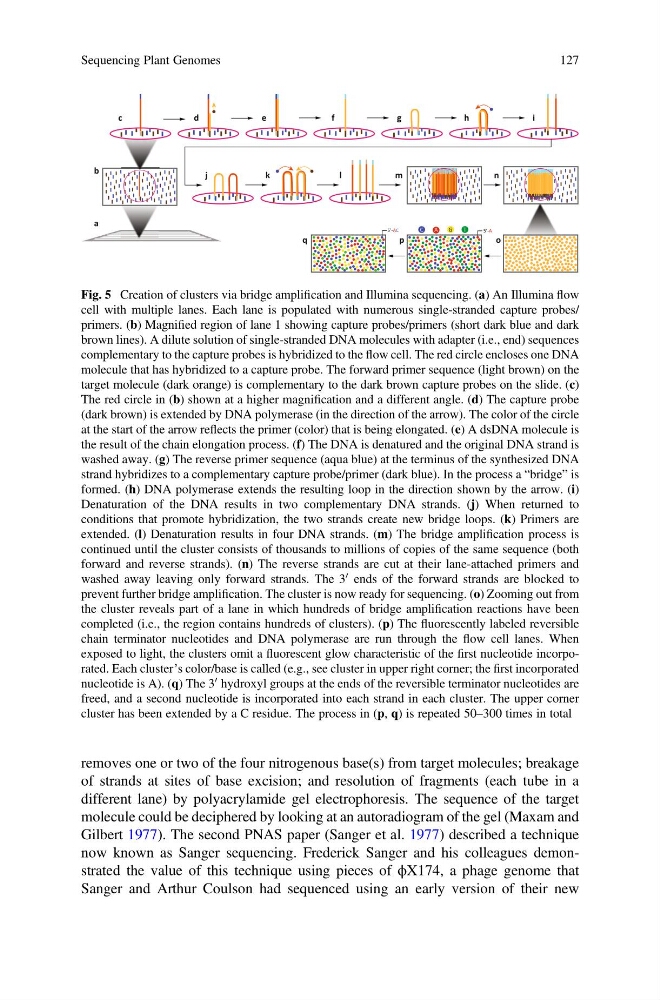
Fig. 5 Creation of clusters via bridge amplification and Illumina sequencing. (a) An Illumina flow
cell with multiple lanes. Each lane is populated with numerous single-stranded capture probes/
primers. (b) Magnified region of lane 1 showing capture probes/primers (short dark blue and dark
brown lines). A dilute solution of single-stranded DNA molecules with adapter (i.e., end) sequences
complementary to the capture probes is hybridized to the flow cell. The red circle encloses one DNA
molecule that has hybridized to a capture probe. The forward primer sequence (light brown) on the
target molecule (dark orange) is complementary to the dark brown capture probes on the slide. (c)
The red circle in (b) shown at a higher magnification and a different angle. (d) The capture probe
(dark brown) is extended by DNA polymerase (in the direction of the arrow). The color of the circle
at the start of the arrow reflects the primer (color) that is being elongated. (e) A dsDNA molecule is
the result of the chain elongation process. (f) The DNA is denatured and the original DNA strand is
washed away. (g) The reverse primer sequence (aqua blue) at the terminus of the synthesized DNA
strand hybridizes to a complementary capture probe/primer (dark blue). In the process a “bridge” is
formed. (h) DNA polymerase extends the resulting loop in the direction shown by the arrow. (i)
Denaturation of the DNA results in two complementary DNA strands. (j) When returned to
conditions that promote hybridization, the two strands create new bridge loops. (k) Primers are
extended. (l) Denaturation results in four DNA strands. (m) The bridge amplification process is
continued until the cluster consists of thousands to millions of copies of the same sequence (both
forward and reverse strands). (n) The reverse strands are cut at their lane-attached primers and
washed away leaving only forward strands. The 3
0 ends of the forward strands are blocked to
prevent further bridge amplification. The cluster is now ready for sequencing. (o) Zooming out from
the cluster reveals part of a lane in which hundreds of bridge amplification reactions have been
completed (i.e., the region contains hundreds of clusters). (p) The fluorescently labeled reversible
chain terminator nucleotides and DNA polymerase are run through the flow cell lanes. When
exposed to light, the clusters omit a fluorescent glow characteristic of the first nucleotide incorporated. Each cluster’s color/base is called (e.g., see cluster in upper right corner; the first incorporated
nucleotide is A). (q) The 3
0 hydroxyl groups at the ends of the reversible terminator nucleotides are
freed, and a second nucleotide is incorporated into each strand in each cluster. The upper corner
cluster has been extended by a C residue. The process in (p, q) is repeated 50–300 times in total
Sequencing Plant Genomes
127
of strands at sites of base excision; and resolution of fragments (each tube in a
different lane) by polyacrylamide gel electrophoresis. The sequence of the target
molecule could be deciphered by looking at an autoradiogram of the gel (Maxam and
Gilbert 1977). The second PNAS paper (Sanger et al. 1977) described a technique
now known as Sanger sequencing. Frederick Sanger and his colleagues demonstrated the value of this technique using pieces of ϕX174, a phage genome that
Sanger and Arthur Coulson had sequenced using an early version of their new
a
b
c
d
e
f
g
h
i
j
k
l
m
n
q
p
o
Fig. 5 Creation of clusters via bridge amplification and Illumina sequencing. (a) An Illumina flow
cell with multiple lanes. Each lane is populated with numerous single-stranded capture probes/
primers. (b) Magnified region of lane 1 showing capture probes/primers (short dark blue and dark
brown lines). A dilute solution of single-stranded DNA molecules with adapter (i.e., end) sequences
complementary to the capture probes is hybridized to the flow cell. The red circle encloses one DNA
molecule that has hybridized to a capture probe. The forward primer sequence (light brown) on the
target molecule (dark orange) is complementary to the dark brown capture probes on the slide. (c)
The red circle in (b) shown at a higher magnification and a different angle. (d) The capture probe
(dark brown) is extended by DNA polymerase (in the direction of the arrow). The color of the circle
at the start of the arrow reflects the primer (color) that is being elongated. (e) A dsDNA molecule is
the result of the chain elongation process. (f) The DNA is denatured and the original DNA strand is
washed away. (g) The reverse primer sequence (aqua blue) at the terminus of the synthesized DNA
strand hybridizes to a complementary capture probe/primer (dark blue). In the process a “bridge” is
formed. (h) DNA polymerase extends the resulting loop in the direction shown by the arrow. (i)
Denaturation of the DNA results in two complementary DNA strands. (j) When returned to
conditions that promote hybridization, the two strands create new bridge loops. (k) Primers are
extended. (l) Denaturation results in four DNA strands. (m) The bridge amplification process is
continued until the cluster consists of thousands to millions of copies of the same sequence (both
forward and reverse strands). (n) The reverse strands are cut at their lane-attached primers and
washed away leaving only forward strands. The 3
0 ends of the forward strands are blocked to
prevent further bridge amplification. The cluster is now ready for sequencing. (o) Zooming out from
the cluster reveals part of a lane in which hundreds of bridge amplification reactions have been
completed (i.e., the region contains hundreds of clusters). (p) The fluorescently labeled reversible
chain terminator nucleotides and DNA polymerase are run through the flow cell lanes. When
exposed to light, the clusters omit a fluorescent glow characteristic of the first nucleotide incorporated. Each cluster’s color/base is called (e.g., see cluster in upper right corner; the first incorporated
nucleotide is A). (q) The 3
0 hydroxyl groups at the ends of the reversible terminator nucleotides are
freed, and a second nucleotide is incorporated into each strand in each cluster. The upper corner
cluster has been extended by a C residue. The process in (p, q) is repeated 50–300 times in total
Sequencing Plant Genomes
127