

g
e
c
b
a
c
d
30 nm
f
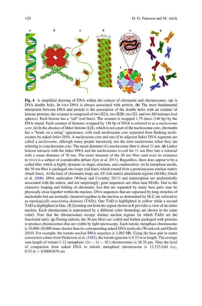
Fig. 4 A simplified drawing of DNA within the context of chromatin and chromosomes. (a) A
DNA double helix. In vivo DNA is always associated with protein. (b) The most fundamental
interaction between DNA and protein is the association of the double helix with an octamer of
histone proteins; the octamer is composed of two H2A, two H2B, two H3, and two H4 histones (red
spheres). Each histone has a “tail” (red lines). The octamer is wrapped 1.75 times (146 bp) by the
DNA strand. Each octamer of histones wrapped by 146 bp of DNA is referred to as a nucleosome
core. (c) In the absence of linker histone (LH), which is not a part of the nucleosome core, chromatin
has a “beads on a string” appearance, with each nucleosome core separated from flanking nucleosomes by naked linker DNA. A nucleosome core and one of its adjacent linker DNA segments are
called a nucleosome, although many people incorrectly use the term nucleosome when they are
referring to a nucleosome core. The mean diameter of a nucleosome fiber is about 11 nm. (d) Linker
histone interacts with the linker DNA and the nucleosomes to coil the 11 nm fiber into a solenoid
with a mean diameter of 30 nm. The exact structure of the 30 nm fiber (and even its existence
in vivo) is a subject of considerable debate (Iyer et al. 2011). Regardless, there does appear to be a
coiled fiber which is highly dynamic in shape, structure, and condensation. (e) In interphase nuclei,
the 30 nm fiber is packaged into loops (red lines) which extend from a proteinaceous nuclear matrix
(black lines). At the base of chromatin loops are AT-rich matrix attachment regions (MARs; Gluch
et al. 2008). DNA replication (Wilson and Coverley 2013) and transcription are preferentially
associated with the matrix, and not surprisingly, gene sequences are often near MARs. Due to the
extensive looping and folding of chromatin, loci that are separated by many base pairs may be
physically close together within the nucleus. DNA sequences that are separated by long stretches of
nucleotides but are normally clustered together in the nucleus as determined by Hi-C are referred to
as topologically associating domains (TADs). One TAD is highlighted in yellow while a second
TAD is highlighted in blue. (f) Zooming out from the region shown in E provides a view of an entire
nucleus. Each chromosome is represented by a different color (homologs are shown in the same
color). Note that the chromosomes occupy distinct nuclear regions (in which TADs are the
functional unit). (g) During mitosis, the 30 nm fibers are coiled and further packaged with proteins
to produce chromosomes that are visible by light microscopy. Each mitotic metaphase chromosome
is 10,000–20,000 times shorter than its corresponding naked DNA molecule (Woodcock and Ghosh
2010). For example, the tomato nuclear DNA sequence is 1,002 Mb. Using the base pair to meter
conversion values from Dickerson et al. (1982), the tomato genome is 0.33 m in length. The average
sum length of tomato’s 12 metaphase (1n ¼ 1x ¼ 1C) chromosomes is 26.76 μm. Thus the level
of compaction from naked DNA to mitotic metaphase chromosome is 12,333-fold (i.e.,
0.33 m Ä 0.00002676 m)
120
D. G. Peterson and M. Arick
e
c
b
a
c
d
30 nm
f
Fig. 4 A simplified drawing of DNA within the context of chromatin and chromosomes. (a) A
DNA double helix. In vivo DNA is always associated with protein. (b) The most fundamental
interaction between DNA and protein is the association of the double helix with an octamer of
histone proteins; the octamer is composed of two H2A, two H2B, two H3, and two H4 histones (red
spheres). Each histone has a “tail” (red lines). The octamer is wrapped 1.75 times (146 bp) by the
DNA strand. Each octamer of histones wrapped by 146 bp of DNA is referred to as a nucleosome
core. (c) In the absence of linker histone (LH), which is not a part of the nucleosome core, chromatin
has a “beads on a string” appearance, with each nucleosome core separated from flanking nucleosomes by naked linker DNA. A nucleosome core and one of its adjacent linker DNA segments are
called a nucleosome, although many people incorrectly use the term nucleosome when they are
referring to a nucleosome core. The mean diameter of a nucleosome fiber is about 11 nm. (d) Linker
histone interacts with the linker DNA and the nucleosomes to coil the 11 nm fiber into a solenoid
with a mean diameter of 30 nm. The exact structure of the 30 nm fiber (and even its existence
in vivo) is a subject of considerable debate (Iyer et al. 2011). Regardless, there does appear to be a
coiled fiber which is highly dynamic in shape, structure, and condensation. (e) In interphase nuclei,
the 30 nm fiber is packaged into loops (red lines) which extend from a proteinaceous nuclear matrix
(black lines). At the base of chromatin loops are AT-rich matrix attachment regions (MARs; Gluch
et al. 2008). DNA replication (Wilson and Coverley 2013) and transcription are preferentially
associated with the matrix, and not surprisingly, gene sequences are often near MARs. Due to the
extensive looping and folding of chromatin, loci that are separated by many base pairs may be
physically close together within the nucleus. DNA sequences that are separated by long stretches of
nucleotides but are normally clustered together in the nucleus as determined by Hi-C are referred to
as topologically associating domains (TADs). One TAD is highlighted in yellow while a second
TAD is highlighted in blue. (f) Zooming out from the region shown in E provides a view of an entire
nucleus. Each chromosome is represented by a different color (homologs are shown in the same
color). Note that the chromosomes occupy distinct nuclear regions (in which TADs are the
functional unit). (g) During mitosis, the 30 nm fibers are coiled and further packaged with proteins
to produce chromosomes that are visible by light microscopy. Each mitotic metaphase chromosome
is 10,000–20,000 times shorter than its corresponding naked DNA molecule (Woodcock and Ghosh
2010). For example, the tomato nuclear DNA sequence is 1,002 Mb. Using the base pair to meter
conversion values from Dickerson et al. (1982), the tomato genome is 0.33 m in length. The average
sum length of tomato’s 12 metaphase (1n ¼ 1x ¼ 1C) chromosomes is 26.76 μm. Thus the level
of compaction from naked DNA to mitotic metaphase chromosome is 12,333-fold (i.e.,
0.33 m Ä 0.00002676 m)
120
D. G. Peterson and M. Arick